Description of the Endothoracic Fascia
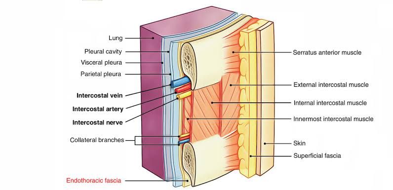
The endothoracic fascia is a significant anatomical structure located within the thoracic cavity. It is defined as a layer of loose connective tissue that lies deep to the intercostal spaces and ribs, effectively separating these structures from the underlying pleura, which is the membrane surrounding the lungs. This fascial layer serves as the outermost membrane of the thoracic cavity and plays a crucial role in providing structural support and facilitating movement between adjacent tissues.
Attachments of Endothoracic Fascia
- Intercostal Spaces: The endothoracic fascia is closely associated with the intercostal muscles, which are situated between adjacent ribs. It provides a supportive layer that allows for the expansion and contraction of these muscles during respiration.
- Ribs: The fascia attaches to the inner surfaces of the ribs, contributing to the overall integrity of the thoracic wall.
- Pleura: The endothoracic fascia separates itself from the parietal pleura, which lines the thoracic cavity and covers the lungs. This separation is essential for maintaining pleural pressure dynamics necessary for lung inflation.
- Suprapleural Membrane: At the apices of the lungs, where it becomes more fibrous, it transitions into what is known as the suprapleural membrane (also referred to as Sibson’s fascia). This area provides additional support to prevent herniation of lung tissue into areas above it.
- Internal Thoracic Artery: The endothoracic fascia also separates structures such as the internal thoracic artery from direct contact with parietal pleura, ensuring vascular structures are protected while still allowing for their functional roles in supplying blood to thoracic tissues.
Overall, this fascial layer not only serves as a physical barrier but also plays an important
Suprapleural Membrane
The suprapleural membrane, also known as Sibson’s fascia, is a significant anatomical structure in human anatomy that serves to cover the apex of each lung. It is a thickening of connective tissue and acts as an extension of the endothoracic fascia, which lies between the parietal pleura and the thoracic cage.
Attachments of the Suprapleural Membrane
- Inferior Attachment: The suprapleural membrane is attached inferiorly to the parietal pleura at the apices of the lungs. This connection allows it to provide support and protection to this region.
- Anterior Attachment: Anteriorly, it connects to the inner border of the first rib and costal cartilage. This attachment plays a crucial role in maintaining structural integrity during respiratory movements.
- Posterior Attachment: Posteriorly, it attaches to the transverse processes of vertebra C7. This connection helps stabilize the membrane against changes in intrathoracic pressure.
- Medial Attachment: Medially, it is connected to the mediastinal pleura, which further integrates it into the thoracic cavity’s overall structure.
- Superior Extension: The suprapleural membrane extends approximately one inch above the superior thoracic aperture, reflecting how the lungs themselves extend higher than the top of the ribcage.
The suprapleural membrane plays a vital role in providing rigidity to the thoracic inlet and helps resist changes in intrathoracic pressure during respiration. It also protects cervical fascia and prevents herniation due to injury.


