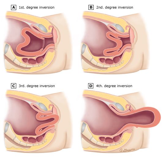
Uterine inversion is a rare but life-threatening obstetric emergency, occurring in approximately 1 in 2,000 to 1 in 20,000 deliveries. It involves the turning of the uterus inside out, where the fundus prolapses through the cervix and may appear at or beyond the vaginal introitus. The condition is most often associated with the third stage of labor and is characterized by a classic triad of postpartum hemorrhage, profound cardiovascular shock, and severe pain. The shock is often disproportionate to the visible blood loss due to a significant neurogenic component (vasovagal shock) triggered by the stretching of peritoneal and pelvic nerves. Prompt recognition and a systematic, multidisciplinary approach are critical to preventing maternal mortality and morbidity.
Immediate Recognition and Call for Help
The first and most crucial step in management is rapid recognition. The clinical presentation is often dramatic and should immediately alert the attending provider.
- Clinical Signs:
- Postpartum Hemorrhage (PPH): Bleeding can range from moderate to torrential.
- Cardiovascular Collapse: The patient may quickly become hypotensive and tachycardic, with signs of shock that seem more severe than the estimated blood loss.
- Severe Abdominal Pain: The patient will often complain of sudden, intense lower abdominal or pelvic pain.
- Visible Mass: A fleshy, dark red or purple mass may be visible at the introitus. This is the inverted uterine fundus. If the placenta is still attached, it will be the leading part of the mass.
- Inability to Palpate the Fundus: An abdominal examination will reveal the absence of the uterine fundus in its expected position. Instead, a dimple or depression may be felt where the fundus should be.
Upon suspicion of uterine inversion, the provider must immediately call for help. This is not a situation that can be managed by a single individual. Announce the emergency clearly (e.g., “Uterine Inversion in Room 5!”) to mobilize a comprehensive team, including additional obstetricians, anesthesiologists, nurses, and personnel to manage blood products. A crucial initial directive is: Do not remove the placenta if it is still attached. Premature removal can trigger catastrophic hemorrhage from the exposed placental implantation site.
Resuscitation and Hemodynamic Stabilization
Resuscitation must occur simultaneously with preparations for uterine replacement. The patient’s life is at immediate risk from hemorrhagic and neurogenic shock.
- Establish IV Access: Secure at least two large-bore (14- or 16-gauge) intravenous cannulas.
- Fluid Resuscitation: Begin aggressive volume replacement with warmed crystalloid solutions (e.g., Lactated Ringer’s or normal saline).
- Activate Massive Transfusion Protocol (MTP): If the patient is in shock or bleeding is massive, activate the MTP without delay. Send blood for type and cross-match, a complete blood count, and coagulation studies. Begin transfusion with O-negative blood if cross-matched blood is not immediately available.
- Oxygenation: Administer high-flow oxygen via a non-rebreather mask to ensure adequate tissue oxygenation.
- Continuous Monitoring: Place the patient on continuous monitoring for heart rate, blood pressure, oxygen saturation, and respiratory rate.
Uterine Relaxation
Before any attempt at repositioning, the uterus and the constricting cervical ring must be relaxed. A tense, contracted uterus is nearly impossible to replace and forceful attempts can cause uterine rupture or increased trauma. The choice of agent depends on availability and the clinical setting.
- Tocolytic Agents:
- Nitroglycerin: This is often a first-line choice due to its rapid onset and short half-life. It can be administered as an intravenous bolus of 50-200 mcg.
- Terbutaline: A beta-mimetic agent, administered as 0.25 mg intravenously or subcutaneously, also provides rapid uterine relaxation.
- Anesthetic Agents: If an anesthesiologist is present, general anesthesia with a halogenated inhalational agent (e.g., sevoflurane, isoflurane) is highly effective at producing profound uterine relaxation. This is often the preferred method if initial attempts with tocolytics fail or if the patient requires transfer to an operating room.
Magnesium sulfate is generally not recommended for this purpose due to its slow onset of action.
Manual Repositioning (The Johnson Maneuver)
Once uterine relaxation is achieved, immediate manual repositioning should be attempted. The Johnson maneuver is the standard technique.
- The operator places a gloved hand into the vagina, cupping the inverted fundus entirely within the palm, with the fingertips at the fornices.
- The fundus is then firmly and steadily pushed upward, through the cervix, along the long axis of the vagina towards the umbilicus.
- The other hand is placed on the maternal abdomen to provide counter-pressure and help guide the uterus back into the abdominal cavity.
- Once the uterus is replaced, the operator’s hand must remain inside the uterine cavity, forming a fist to act as an internal tamponade and maintain fundal pressure. This prevents immediate re-inversion.
- While the hand is in place, the team should immediately begin a high-dose oxytocin infusion to stimulate uterine contraction.
- The internal hand should only be withdrawn after the uterus has become firm and contracted around the fist and all uterotonic agents have been administered.
If this maneuver fails, a hydrostatic method can be attempted before proceeding to surgery. The O’Sullivan method involves instilling 2-5 liters of warm saline into the vagina while the labia are manually sealed, using the fluid pressure to push the fundus back into place.
Post-Repositioning Management and Prevention of Recurrence
Replacing the uterus is only half the battle; preventing its recurrence is equally vital.
- Administer Uterotonics: A multi-agent approach is recommended to ensure a firm, sustained uterine contraction.
- Oxytocin: A high-dose infusion (e.g., 20-40 units in 1 liter of crystalloid) should be running.
- Methylergonovine (Methergine): 0.2 mg IM (contraindicated in patients with hypertension).
- Carboprost Tromethamine (Hemabate): 250 mcg IM (contraindicated in patients with asthma).
- Misoprostol: 800-1000 mcg administered rectally.
- Placental Removal: If the placenta was left attached, it can now be manually removed once the uterus is repositioned and firm.
- Bimanual Compression: Continue bimanual uterine compression until the uterus is well-contracted and bleeding has ceased.
- Intensive Monitoring: The patient requires close observation in a high-dependency setting for at least 24 hours. Monitor vital signs, uterine tone, and vaginal bleeding frequently. Continue the oxytocin infusion for several hours.
- Antibiotics: Consider broad-spectrum prophylactic antibiotics, as the exposed endometrial cavity is at high risk for infection.
Surgical Management
If manual and hydrostatic methods fail, surgical intervention is required. This is typically performed via laparotomy in an operating room.
- Huntington Procedure: This is the primary surgical technique. After performing a laparotomy, the inverted fundus can be seen as a cup-like depression in the pelvis. The surgeon grasps the round ligaments and the uterine wall within the “cup” with Allis or Babcock clamps and applies gentle, steady, upward traction. This is done sequentially on each side, gradually pulling the uterus out of its inversion.
- Haultain Procedure: This is used if the Huntington procedure fails, usually due to an extremely tight and fibrotic constriction ring. A vertical incision is made posteriorly through the constriction ring on the inverted uterus. This releases the tension, allowing the fundus to be repositioned, after which the uterine incision is repaired in layers.
In summary, the management of uterine inversion is a time-critical emergency that follows a logical sequence: Recognize, Resuscitate, Relax, Replace, and Reinforce with uterotonics. A calm, coordinated, and multidisciplinary team approach is paramount to achieving a successful maternal outcome.
References
- American College of Obstetricians and Gynecologists. (2017). ACOG Practice Bulletin No. 183: Postpartum Hemorrhage. Obstetrics & Gynecology, 130(4), e168-e186.
- Cunningham, F. G., Leveno, K. J., Bloom, S. L., Dashe, J. S., Hoffman, B. L., Casey, B. M., & Spong, C. Y. (Eds.). (2018). Williams Obstetrics (25th ed.). McGraw-Hill Education.
- Gandhi, A., Guntupalli, S. R., & Raga, F. (2021). Puerperal Uterine Inversion. In UpToDate. Retrieved from https://www.uptodate.com
- Witteveen, T., van Stralen, G., Zwart, J., & van Roosmalen, J. (2013). Puerperal uterine inversion in the Netherlands: a nationwide cohort study. Acta Obstetricia et Gynecologica Scandinavica, 92(3), 334-340.
- Shellhaas, C. S. (2020). The management of uterine inversion. Contemporary OB/GYN, 65(1).


