Epithelial tissue, a fundamental component of the human body, forms a protective barrier, lines cavities, and is involved in secretion and absorption. Understanding its diverse forms is crucial for histology and pathology.
Defining Epithelial Tissue
Epithelial tissue, also known as epithelium, is characterized by its closely packed cells that form continuous sheets. These cells are arranged on an extracellular matrix called the basement membrane, which separates the epithelium from the underlying connective tissue. Key features of epithelial tissue include:
- Cellularity: Epithelial tissues are composed almost entirely of cells with very little extracellular material.
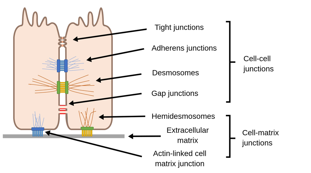
- Specialized Contacts: Cells within an epithelium are joined by specialized junctions, such as tight junctions, adherens junctions, desmosomes, and gap junctions, which maintain the integrity of the tissue.
- Polarity: Epithelial cells typically exhibit polarity, meaning they have an apical (free or exposed) surface and a basal (attached) surface. The apical surface may possess modifications like microvilli or cilia, while the basal surface is anchored to the basement membrane.
- Supported by Connective Tissue: All epithelial sheets are supported by an underlying layer of connective tissue, which provides nourishment and structural support.
- Avascular but Innervated: Epithelial tissues are avascular, meaning they lack blood vessels. They receive nutrients via diffusion from the underlying connective tissue. However, they are innervated, containing nerve endings.
- Regenerative Capacity: Epithelial tissues have a high capacity for regeneration and repair, replacing damaged cells rapidly.
Classification of Epithelial Tissues
Epithelial tissues are classified based on two primary criteria: the number of cell layers and the shape of the cells.
1. Classification Based on Number of Cell Layers:
- Simple Epithelium: Consists of a single layer of cells. These epithelia are typically found where absorption, secretion, and filtration occur, as the thinness of the tissue facilitates these processes.
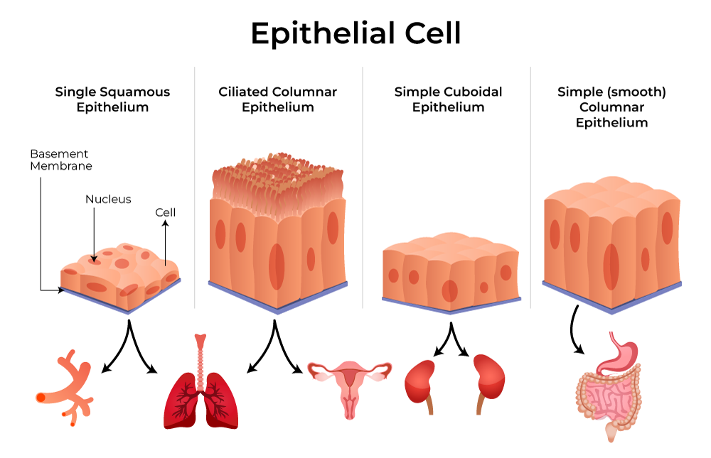
- Simple Squamous Epithelium: Characterized by a single layer of flattened, scale-like cells with a centrally flattened nucleus. The cytoplasm is sparse.
- Function: Primarily involved in diffusion and filtration. Its thinness makes it ideal for exchange.
- Locations: Found in the lining of blood vessels (endothelium), lymphatic vessels, serous membranes (mesothelium), alveoli of the lungs, and Bowman’s capsule of the kidney.
- Microscopic Appearance: Appears as a thin, continuous sheet of flattened cells, often with a somewhat irregular shape. The nuclei are visible as flattened discs. In cross-section, it forms a very thin barrier.
- Simple Cuboidal Epithelium: Composed of a single layer of cube-shaped cells that are approximately as tall as they are wide. The nuclei are typically spherical and centrally located.
- Function: Involved in secretion and absorption.
- Locations: Found in the kidney tubules, ducts of glands (e.g., salivary glands, pancreas), and the surface of the ovary.
- Microscopic Appearance: Cells present a square or rectangular profile in cross-section. The lumen (internal space) of ducts or tubules is usually round. The nuclei are prominent and spherical.
- Simple Columnar Epithelium: Consists of a single layer of tall, column-shaped cells. The nuclei are typically elongated and located near the base of the cells. The apical surface may have specializations like microvilli or cilia.
- Function: Primarily involved in absorption and secretion. Modifications like microvilli increase surface area for absorption, while goblet cells within the epithelium secrete mucus. Ciliated variants propel substances.
- Locations: Found lining the digestive tract from the stomach to the rectum, the gallbladder, and the uterine tubes. Ciliated simple columnar epithelium lines the uterine tubes and parts of the respiratory tract.
- Microscopic Appearance: Cells are significantly taller than they are wide, appearing rectangular in cross-section. The nuclei are oval and basally located. If microvilli are present, the apical surface may appear fuzzy (brush border). Goblet cells, which appear as clear, goblet-shaped cells filled with mucus, are often interspersed.
- Pseudostratified Columnar Epithelium: Although appearing to have multiple layers due to the varying heights of the nuclei, all cells in this epithelium actually rest on the basement membrane. However, not all reach the apical surface. It is often ciliated.
- Function: Primarily involved in secretion and propulsion of mucus.
- Locations: Most common in the lining of the trachea and the upper respiratory tract. Also found in ducts of large glands and parts of the male reproductive tract.
- Microscopic Appearance: This is a distinctive type. The nuclei are at different levels, giving the impression of stratification. However, a careful examination reveals that all cell bases are connected to the basement membrane. The apical surface is often covered with cilia. Goblet cells are frequently present.
- Simple Squamous Epithelium: Characterized by a single layer of flattened, scale-like cells with a centrally flattened nucleus. The cytoplasm is sparse.
- Stratified Epithelium: Composed of two or more layers of cells. These epithelia are found in areas subjected to abrasion and stress, offering protection.
- Stratified Squamous Epithelium: The most widespread stratified epithelium. The apical cells are flattened and scale-like, while the basal cells are more cuboidal or columnar.
- Function: Primarily protective.
- Types:
- Keratinized Stratified Squamous Epithelium: The apical layers of cells are dead, flattened cells that have filled with keratin. This forms a tough, waterproof outer layer.
- Locations: Forms the epidermis of the skin.
- Microscopic Appearance: Distinct layers are visible. The superficial cells are thin, anucleated, and densely packed with keratin, appearing pinkish (eosinophilic) and often flaky. Deeper layers have increasingly viable cells, with basal cells being actively mitotic.
- Non-keratinized Stratified Squamous Epithelium: The apical cells are flattened but possess visible nuclei and are moist. They do not contain significant amounts of keratin.
- Locations: Lines the moist internal surfaces such as the lining of the mouth, esophagus, vagina, and anal canal.
- Microscopic Appearance: Similar to keratinized, but the surface cells are nucleated and appear less eosinophilic and not as flattened.
- Keratinized Stratified Squamous Epithelium: The apical layers of cells are dead, flattened cells that have filled with keratin. This forms a tough, waterproof outer layer.
- Stratified Cuboidal Epithelium: Typically consists of two layers of cuboidal cells.
- Function: Involved in protection and secretion.
- Locations: Relatively rare. Found in the ducts of sweat glands, mammary glands, and salivary glands.
- Microscopic Appearance: Appears as multiple layers of roughly square-shaped cells. The lumen of the duct is often small.
- Stratified Columnar Epithelium: Consists of multiple layers of cells, with the apical layer being columnar and the basal layers typically cuboidal.
- Function: Involved in protection and secretion.
- Locations: Limited distribution. Found in small amounts in the pharynx, male urethra, and lining of some glandular ducts.
- Microscopic Appearance: The surface cells are distinctly columnar, while the underlying cells are smaller and more irregularly shaped.
- Transitional Epithelium: A specialized stratified epithelium found only in the urinary system. It is characterized by its ability to change shape, allowing organs to stretch. The basal cells are cuboidal or columnar, while the apical cells are large and dome-shaped or squamous-like, depending on whether the organ is stretched or relaxed.
- Function: Permits distension of urinary organs.
- Locations: Lines the ureters, urinary bladder, and proximal urethra.
- Microscopic Appearance: When the organ is relaxed, the apical cells are large and rounded (umbrella cells). When the organ is stretched, these cells flatten significantly, and the epithelium appears thinner. The stratified nature is evident, with multiple layers.
- Stratified Squamous Epithelium: The most widespread stratified epithelium. The apical cells are flattened and scale-like, while the basal cells are more cuboidal or columnar.
2. Classification Based on Cell Shape:
As described above, the shape of the cells also plays a crucial role in classification:
- Squamous Cells: Flattened, irregular shape, like scales. Their nuclei are flattened discs.
- Cuboidal Cells: Cube-shaped, approximately as tall as they are wide. Their nuclei are spherical.
- Columnar Cells: Tall and rectangular, taller than they are wide. Their nuclei are elongated and usually basally located.
Specialized Epithelial Modifications:
Beyond the basic classification, epithelial cells can exhibit specialized structures on their apical surface that enhance their function:
- Microvilli: Finger-like projections of the plasma membrane that increase surface area for absorption. Found in the lining of the small intestine and kidney tubules. Appears as a “brush border” under the microscope.
- Cilia: Hair-like projections that beat in a coordinated manner to move substances along the surface. Found in the respiratory tract (to move mucus) and uterine tubes (to move eggs). Appear as short, motile projections.
- Stereocilia: Very long, non-motile microvilli that resemble cilia but are much longer and branched. Found in the epididymis and inner ear.
Summary Table for Light Microscope Identification:
| Epithelial Type | Number of Layers | Cell Shape (Apical) | Key Features/Locations |
|---|---|---|---|
| Simple Squamous | One | Flattened | Thin barrier; endothelium, mesothelium, alveoli, kidney tubules |
| Simple Cuboidal | One | Cube-shaped | Secretion/absorption; kidney tubules, gland ducts, ovary |
| Simple Columnar | One | Columnar | Absorption/secretion; digestive tract, gall bladder, uterine tubes |
| Pseudostratified Columnar | Appears many, is one | Columnar (varied) | Often ciliated; trachea, respiratory tract, male reproductive tract |
| Stratified Squamous (Keratinized) | Many | Squamous (keratinized) | Protection; epidermis of skin |
| Stratified Squamous (Non-keratinized) | Many | Squamous (nucleated) | Protection; mouth, esophagus, vagina, anal canal |
| Stratified Cuboidal | Two or more | Cuboidal | Protection/secretion; ducts of sweat/mammary/salivary glands |
| Stratified Columnar | Many | Columnar | Protection/secretion; pharynx, male urethra, large gland ducts |
| Transitional | Many (specialized) | Dome-shaped/squamous | Stretches; urinary bladder, ureters, proximal urethra |
Conclusion
The diverse array of epithelial tissues, categorized by the number of cell layers and the shape of the cells, plays vital roles in maintaining homeostasis and protecting the body. By carefully observing the cellular arrangement, nuclear morphology, and any apical specializations under a light microscope, one can accurately identify and understand the function of these fundamental tissues, forming a critical basis for further biological and medical studies.
References:
- Alberts, B., Johnson, A., Lewis, J., Raff, M., Roberts, K., & Walter, P. (2015). Molecular Biology of the Cell (6th ed.). Garland Science.
- Kierszenbaum, A. L., & Tres, L. L. (2015). Histology and Cell Biology: An Introduction to Pathology (4th ed.). Elsevier.
- Mescher, A. L., & Junqueira, L. C. (2013). Junqueira’s Basic Histology: Text and Atlas (13th ed.). McGraw-Hill Education.
- Ross, M. H., & Pawlina, W. (2016). Histology: A Text and Atlas: With Correlated Cell and Molecular Biology (7th ed.). Wolters Kluwer.
- Widmaier, E. P., Raff, H., & Strang, K. (2016). Vander’s Human Physiology: The Mechanisms of Body Function (14th ed.). McGraw-Hill Education.


