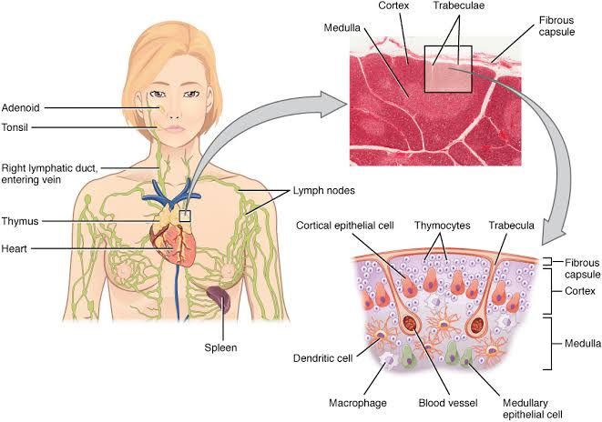
The lymphatic system is a vital component of the immune system, comprising a network of vessels, tissues, and organs that help rid the body of toxins, waste, and other unwanted materials. Histological examination under a light microscope is a fundamental method for identifying and characterizing these organs, understanding their structure, and correlating structure with function.
General Principles of Histological Examination
Before examining specific organs, it is helpful to understand the general criteria common to many lymphoid tissues under the microscope:
- High Cellularity: Lymphoid organs are typically rich in cells, predominantly lymphocytes.
- Presence of Lymphocytes: While other cell types are present, lymphocytes (small, round, darkly stained nuclei with scant cytoplasm) are a defining feature.
- Connective Tissue Stroma: A framework of connective tissue, often including reticular fibers, supports the parenchyma (functional tissue). This may include a capsule, trabeculae, and a reticular meshwork.
- Vascular Supply: Lymphoid organs are well-vascularized.
- Lymphoid Aggregations (Follicles): Collections of B lymphocytes, often organized into primary (uniform small lymphocytes) and secondary (germinal center with larger, actively dividing cells) follicles. Note that not all lymphoid organs contain follicles (e.g., thymus).
Examination and Identification of Lymphoid Organs
The following steps outline the process for examining prepared histological slides of lymphoid organs. Begin each examination at low power, scanning the entire cross-section to grasp the overall architecture before moving to higher magnifications for detailed analysis.
Organ 1: Lymph Node
Lymph nodes are small, encapsulated organs strategically located along lymphatic vessels. They filter lymph and serve as sites for initiating immune responses.
- Step 1: Low Power Scan (e.g., 4x or 10x objective)
- Overall Shape and Capsule: Identify the outer connective tissue capsule, typically a dense fibrous band. Note the overall bean or kidney shape, with a convex outer surface and a concave hilum where blood vessels enter and leave.
- Trabeculae: Observe extensions of the capsule (trabeculae) penetrating into the interior of the node.
- Gross Regional Differentiation: Look for a distinct organization into an outer cortex and an inner medulla. The cortex appears denser and often contains spherical structures, while the medulla appears lighter and cord-like.
- Step 2: Medium Power Examination (e.g., 10x or 20x objective) – Cortex
- Subcapsular Sinus: Directly beneath the capsule, identify the subcapsular (marginal) sinus, a clear space where afferent lymphatic vessels empty.
- Lymphoid Follicles: Within the cortex, observe the characteristic lymphoid follicles. These are roughly spherical aggregates.
- Primary Follicles: Composed uniformly of small, darkly stained lymphocytes.
- Secondary Follicles: Contain a lighter-staining central region (germinal center), surrounded by a darker mantle zone (corona) of small lymphocytes. Germinal centers are sites of B cell proliferation and differentiation.
- Paracortex: Located between the cortex and medulla, this region is primarily populated by T lymphocytes. It appears less densely packed than the cortex and lacks follicles. Identify high endothelial venules (HEVs) within the paracortex – specialized blood vessels with plump (cuboidal or columnar) endothelial cells, crucial for lymphocyte entry from the blood.
- Step 3: Medium/High Power Examination (e.g., 20x or 40x objective) – Medulla
- Medullary Cords: Observe irregular cords of lymphoid tissue extending from the paracortex. These cords contain lymphocytes (including plasma cells), macrophages, and dendritic cells.
- Medullary Sinuses: Identify the irregular, endothelium-lined spaces separating the medullary cords. These sinuses converge towards the hilum and contain lymph flowing towards the efferent lymphatic vessel. Macrophages are often visible lining the sinuses.
- Step 4: Identify Key Distinguishing Features of Lymph Node
- Presence of a distinct capsule.
- Clear zonation into cortex and medulla.
- Presence of B cell follicles (primary and/or secondary) predominantly in the cortex.
- Presence of a paracortex rich in T cells and HEVs.
- Structure of medulla as cords separated by sinuses.
- Presence of subcapsular, cortical, and medullary sinuses containing filtering lymph.
Organ 2: Spleen
The spleen is the largest lymphoid organ, located in the abdomen. It filters blood, removes old or damaged red blood cells, and initiates immune responses to bloodborne antigens.
- Step 1: Low Power Scan (e.g., 4x or 10x objective)
- Overall Shape and Capsule: Identify the outer capsule, a dense connective tissue layer containing smooth muscle (variable by species). Trabeculae extend deep into the parenchyma.
- Absence of Cortex/Medulla: Unlike the lymph node, there is no distinct cortex-medulla segregation in the same sense. Instead, the parenchyma is divided into red pulp and white pulp.
- Distribution of Red and White Pulp: At low power, identify areas of white pulp as discrete, pale-staining islands or nodules embedded within the more extensive, darker-staining red pulp.
- Step 2: Medium Power Examination (e.g., 10x or 20x objective) – White Pulp
- Central Arteriole: A key identifying feature. Locate the small arteriole, often eccentrically placed, within each aggregate of white pulp. This is a branch of the splenic artery.
- Periarteriolar Lymphoid Sheath (PALS): The cuff of lymphocytes (primarily T cells) surrounding the central arteriole.
- Splenic Follicles: Often attached to the PALS, these are aggregates of B cells, structurally similar to lymph node follicles, frequently containing prominent germinal centers.
- Marginal Zone: A less densely cellular region surrounding the white pulp, separating it from the red pulp. It contains specialized macrophages, B cells, and dendritic cells involved in capturing bloodborne antigens.
- Step 3: Medium/High Power Examination (e.g., 20x or 40x objective) – Red Pulp
- Splenic Cords (of Billroth): Identify the anastomosing cords of connective tissue containing a variety of cells, including erythrocytes, macrophages, lymphocytes, plasma cells, and granulocytes. This is the primary site for filtering blood.
- Splenic Sinusoids: Observe the irregular, wide vascular channels separating the splenic cords. Look for the characteristic stave cells (elongated endothelial cells) and hoops of reticular fibers that comprise the sinusoid walls, especially visible with special stains (though not always required for basic identification). Note the abundance of red blood cells within the sinusoids and cords.
- Step 4: Identify Key Distinguishing Features of Spleen
- Presence of a capsule and prominent trabeculae extending throughout the parenchyma.
- Lack of distinct cortex/medulla zonation like the lymph node.
- Division into morphologically distinct red pulp and white pulp.
- Presence of a central arteriole within the white pulp (a defining feature, unlike lymph nodes).
- Structure of white pulp including PALS and splenic follicles.
- Structure of red pulp including splenic cords and sinusoids containing abundant erythrocytes.
Organ 3: Thymus
The thymus is a primary lymphoid organ located in the mediastinum. It is essential for the development and maturation of T lymphocytes (T cells). It is most active during childhood and undergoes involution (shrinks) after puberty.
- Step 1: Low Power Scan (e.g., 4x or 10x objective)
- Overall Structure: Note the lobulated appearance. The organ is divided into numerous lobules by connective tissue septa (trabeculae) extending from the capsule.
- Regional Differentiation within Lobules: Each lobule shows a distinct darker-staining outer cortex and a lighter-staining inner medulla.
- Step 2: Medium Power Examination (e.g., 10x or 20x objective) – Cortex
- Dense Cellularity: The cortex is very densely packed with small, darkly staining cells – developing T lymphocytes or thymocytes.
- Epithelial Reticular Cells: Identify the larger, pale-staining nuclei and eosinophilic cytoplasm of the epithelial reticular cells, which form a supportive network. They are less easily seen amongst the dense thymocytes at this magnification.
- Absence of Follicles: Crucially, there are no lymphoid follicles in the thymus, distinguishing it from lymph nodes, spleen, and tonsils.
- Step 3: Medium Power/High Power Examination (e.g., 20x or 40x objective) – Medulla
- Lower Cellularity: The medulla is less densely packed with lymphocytes than the cortex.
- Prominent Epithelial Reticular Cells: These cells are more apparent in the medulla.
- Hassall’s Corpuscles: The most characteristic feature of the thymic medulla. Identify eosinophilic (pink), concentric layers of flattened epithelial reticular cells. Their size and appearance can vary, and they may sometimes show signs of keratinization or calcification. These are unique to the thymus.
- Step 4: Identify Key Distinguishing Features of Thymus
- Distinct lobulated architecture.
- Clear zonation into cortex and medulla within each lobule.
- Absence of lymphoid follicles.
- Presence of Hassall’s corpuscles in the medulla (a definitive feature).
- Stroma composed of epithelial reticular cells, not reticular fibers (though both are present, the epithelial component is key).
- High density of thymocytes in the cortex.
- Absence of afferent lymphatic vessels (lymphoid cells enter via blood, not lymph).
Organ 4: Tonsils
Tonsils are unencapsulated or partially encapsulated lymphoid tissues located in the wall of the pharynx. They monitor antigens entering the body via the oral and nasal cavities.
- Step 1: Low Power Scan (e.g., 4x or 10x objective)
- Location and Epithelium: Note the location directly beneath or associated with the epithelial lining (usually stratified squamous or pseudostratified columnar, depending on the specific tonsil).
- Absence of Complete Capsule: Observe the lack of a well-defined, complete connective tissue capsule surrounding the entire organ.
- Presence of Crypts: Identify characteristic deep invaginations of the surface epithelium into the underlying lymphoid tissue. These “crypts” increase the surface area for encountering antigens.
- Step 2: Medium Power Examination (e.g., 10x or 20x objective) – Lymphoid Tissue
- Lymphoid Follicles: Identify numerous lymphoid follicles, often with prominent germinal centers, located beneath or around the crypts. These are similar in appearance to those in lymph nodes and the spleen.
- Diffuse Lymphocytes: Observe lymphocytes scattered diffusely in the connective tissue between the follicles.
- Step 3: High Power Examination (e.g., 20x or 40x objective)
- Epithelial Lining: Examine the surface epithelium and the epithelium lining the crypts. Note that this epithelium is often infiltrated by lymphocytes.
- Follicle Detail: Examine the germinal centers for larger lymphocytes (centroblasts, centrocytes), macrophages, and follicular dendritic cells.
- Step 4: Identify Key Distinguishing Features of Tonsils
- Located beneath or associated with a surface epithelium.
- Presence of characteristic epithelial crypts (especially palatine and lingual tonsils).
- Lack of a complete capsule.
- Presence of lymphoid follicles (often with germinal centers) associated with the epithelium.
Organ 5: Mucosa-Associated Lymphoid Tissue (MALT)
MALT is lymphoid tissue found in the lamina propria and submucosa of mucosal tracts (gastrointestinal, respiratory, genitourinary). It is the largest mass of lymphoid tissue in the body. Examples include Peyer’s patches in the small intestine, lymphoid tissue in the appendix, and bronchial-associated lymphoid tissue (BALT).
- Step 1: Low Power Scan (e.g., 4x or 10x objective)
- Location: Identify the mucosal lining (epithelium, lamina propria, muscularis mucosae) of the specific organ (e.g., intestine, bronchus). The MALT will be located within the lamina propria and/or submucosa.
- Lack of Capsule: Note the absence of a surrounding connective tissue capsule.
- Aggregation: Observe the clustering of lymphoid tissue, often as discrete nodules or a more diffuse infiltration. In structures like Peyer’s patches, this is a prominent aggregation of many follicles.
- Step 2: Medium Power Examination (e.g., 10x or 20x objective)
- Lymphoid Follicles: Identify lymphoid follicles, frequently with prominent germinal centers, embedded within the lamina propria or submucosa. These are structurally similar to other lymphoid follicles.
- Diffuse Lymphocytes: Note the significant population of scattered lymphocytes, plasma cells, and other immune cells throughout the lamina propria surrounding the follicles.
- Association with Epithelium: Observe the close association of the lymphoid tissue with the overlying mucosal epithelium. In specific MALT regions like Peyer’s patches, note the presence of specialized follicle-associated epithelium (containing M cells), although identifying M cells specifically may require higher magnification or special techniques.
- Step 3: High Power Examination (e.g., 20x or 40x objective)
- Cellular Detail: Examine the cell types within the follicles and diffuse areas (lymphocytes, plasma cells, macrophages, etc.).
- Epithelial Interface: Observe the interface between the lymphoid tissue and the mucosal epithelium.
- Step 4: Identify Key Distinguishing Features of MALT
- Located within the lamina propria and/or submucosa of a mucosal organ.
- Lack of a surrounding connective tissue capsule.
- Close association with mucosal epithelium.
- Presence of lymphoid follicles (in organized MALT like Peyer’s patches) and/or diffuse lymphoid infiltration.
- In structures like Peyer’s patches, a prominent aggregation of multiple follicles forming a plaque.
Conclusion
Systematic histological examination, starting at low power to assess overall architecture and progressively increasing magnification to observe cellular detail and specific structures, is essential for accurate identification of lymphoid organs. By focusing on key distinguishing features such as the presence or absence of a capsule, internal zonation (cortex/medulla vs. red/white pulp vs. lobules) the presence or absence and organization of follicles, and the presence of unique structures (Hassall’s corpuscles, central arterioles, crypts, associated epithelium), one can reliably differentiate between lymph nodes, spleen, thymus, tonsils, and MALT. Mastering the identification of these structures is crucial for understanding the functional anatomy of the immune system.


