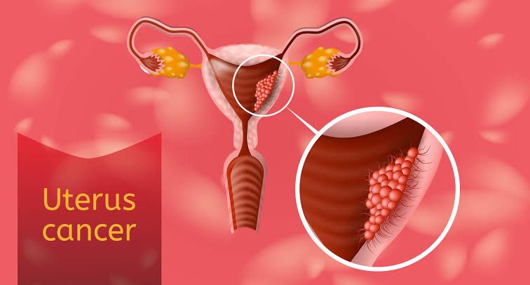
Uterine cancer, also known as endometrial cancer in the vast majority of cases, is a malignancy that arises from the cells of the uterus. It is a common gynecological cancer, particularly affecting women after menopause. Understanding the different types, risk factors, symptoms, diagnostic procedures, prognosis, and management options is crucial for both healthcare professionals and the public. This educational overview provides a structured look at these key aspects.
Types of Uterine Cancer
Uterine cancers are broadly classified based on the tissue from which they originate. The most common type arises from the endometrium, the inner lining of the uterus, while less common types arise from the muscular or connective tissues.
- Endometrial Carcinoma (originating from the endometrium): This accounts for over 90% of uterine cancers. Endometrial carcinomas are further classified based on their microscopic appearance and biological behavior:
- Endometrioid Adenocarcinoma: This is the most common subtype of endometrial cancer, making up 70-80% of cases. It typically arises from endometrial hyperplasia (abnormal thickening of the lining) and is often associated with exposure to estrogen. These are frequently graded based on their differentiation (how closely they resemble normal endometrial cells):
- Grade 1 and 2: Generally considered lower grade, less aggressive, and often associated with a better prognosis, particularly when detected early.
- Grade 3: Considered higher grade, more aggressive, and has a higher risk of recurrence and spread.
- Serous Carcinoma: This is a less common but more aggressive subtype, accounting for about 5-10% of cases. It often arises spontaneously, without association with estrogen or hyperplasia, and tends to be diagnosed at more advanced stages. It has a higher propensity for spread within the abdomen (peritoneal dissemination) and to lymph nodes.
- Clear Cell Carcinoma: Another less common and aggressive subtype, comprising less than 5% of cases. Similar to serous carcinoma, it is not typically linked to estrogen and has a guarded prognosis, often recurring or metastasizing.
- Mucinous Adenocarcinoma: A rare subtype (less than 5%), which can range from low-grade to high-grade depending on the proportion of malignant cells.
- Mixed Cell Carcinoma: Contains a mixture of two or more subtypes, with the prognosis often dictated by the most aggressive component (e.g., a mixture of endometrioid and serous components would be treated based on the serous element).
- Undifferentiated Carcinoma: A rare, highly aggressive type where the cells are so poorly differentiated that they cannot be classified into a specific subtype. It carries a poor prognosis.
- Endometrioid Adenocarcinoma: This is the most common subtype of endometrial cancer, making up 70-80% of cases. It typically arises from endometrial hyperplasia (abnormal thickening of the lining) and is often associated with exposure to estrogen. These are frequently graded based on their differentiation (how closely they resemble normal endometrial cells):
- Uterine Sarcoma (originating from the muscle or connective tissue of the uterus): These are much less common, representing about 2-5% of uterine malignancies. They are generally more aggressive and have a higher risk of recurrence and distant spread than typical endometrial carcinomas.
- Leiomyosarcoma: Arises from the smooth muscle of the uterine wall (myometrium). It can be difficult to distinguish from a benign fibroid (leiomyoma) before surgery. It is the most common type of uterine sarcoma.
- Endometrial Stromal Sarcoma: Originates from the connective tissue (stroma) of the endometrium. These are further divided:
- Low-Grade Endometrial Stromal Sarcoma: Grows more slowly and has a better prognosis than high-grade types or leiomyosarcoma, but can recur late.
- High-Grade Endometrial Stromal Sarcoma: More aggressive with a poorer prognosis.
- Undifferentiated Uterine Sarcoma: A rare, highly aggressive sarcoma with no clear differentiation.
- Carcinosarcoma (also known as Malignant Mixed Mullerian Tumor – MMMT): Represents a rare (about 1%), but highly aggressive, type. Despite having both carcinomatous (epithelial) and sarcomatous (mesenchymal) components, they are now generally considered high-grade epithelial tumors with a sarcomatous transformation, and they behave more like high-grade carcinomas (like serous carcinoma).
Risk Factors
Several factors can increase a woman’s risk of developing uterine cancer, particularly endometrial carcinoma. These often relate to hormonal influences, specifically prolonged or unopposed exposure to estrogen.
- Age: The risk significantly increases with age, with most cases occurring after menopause (typically over 50).
- Obesity: Fat tissue produces estrogen. Higher body mass index (BMI) leads to increased estrogen levels, increasing risk.
- Hormone Replacement Therapy (HRT): Using estrogen-only HRT in women with an intact uterus significantly increases the risk. Combining estrogen with a progestin (combined HRT) lowers this risk, though it may still be slightly elevated compared to no HRT.
- Tamoxifen: A drug used to treat breast cancer. While it blocks estrogen in breast tissue, it acts like estrogen in the uterus, increasing the risk of endometrial cancer (especially low-grade types), although the benefits for breast cancer treatment usually outweigh this risk.
- Reproductive History:
- Never having been pregnant (nulliparity).
- Starting menstruation at an early age (before 12).
- Having menopause at a late age (after 55).
- These factors increase lifetime exposure to estrogen.
- Polycystic Ovary Syndrome (PCOS): This condition can cause irregular or absent ovulation, leading to prolonged exposure of the endometrium to estrogen without the cyclical shedding induced by progesterone, increasing risk.
- Diabetes: Women with diabetes, particularly type 2, have an increased risk. The exact mechanism is not fully understood but may relate to insulin resistance and hormonal imbalances.
- Genetic Syndromes:
- Lynch Syndrome (HNPCC – Hereditary Nonpolyposis Colorectal Cancer): This is the most common genetic predisposition for endometrial cancer. Women with Lynch syndrome have a significantly increased lifetime risk (up to 60%). It is also associated with colorectal, ovarian, and other cancers.
- Previous Pelvic Radiation Therapy: Radiation to the pelvis for other cancers can damage cells and increase the risk of later developing uterine cancer, including sarcomas.
- Family History: Having a first-degree relative (mother, sister, daughter) with endometrial cancer, especially if diagnosed at a young age, can slightly increase risk, even in the absence of a known syndrome like Lynch.
Clinical Presentation
The most common symptom of uterine cancer, particularly endometrial carcinoma, is abnormal vaginal bleeding. However, symptoms can vary and may be absent in early stages.
- Abnormal Vaginal Bleeding:
- Post-menopausal bleeding: Any bleeding, spotting, or staining after menopause is the most significant warning sign and warrants immediate investigation.
- Pre-menopausal or peri-menopausal bleeding: Heavy or prolonged menstrual periods, or bleeding between periods (intermenstrual bleeding).
- Abnormal Vaginal Discharge: A watery, bloody, or foul-smelling discharge, potentially containing pus or blood.
- Pelvic Pain or Pressure: Pain or a feeling of pressure, fullness, or cramping in the lower abdomen or pelvis. This can occur as the tumor grows larger or if it begins to press on nearby structures.
- Pain During Intercourse (Dyspareunia).
- Changes in Bowel or Bladder Habits: Difficulty urinating, pain during urination, or constipation, which can occur if the cancer spreads to press on the bladder or rectum (usually in more advanced stages).
- Unexplained Weight Loss: Can be a symptom of more advanced disease.
- Palpable Abdominal Mass: Less common, may indicate a large tumor (more likely with sarcomas) or advanced disease.
Important Note: Uterine sarcomas may present with similar symptoms (abnormal bleeding or pain) but can also present with a rapidly enlarging uterine mass (previously thought to be a fibroid) or be asymptomatic until late stages.
Appropriate Investigations
If uterine cancer is suspected based on symptoms or risk factors, a series of investigations are performed to confirm the diagnosis, determine the type and extent of the cancer, and plan treatment.
- Medical History and Physical Examination: Detailed history of symptoms, risk factors, and general health. Pelvic examination to assess the size and shape of the uterus and check for any masses.
- Endometrial Biopsy: This is the primary diagnostic test for suspected endometrial cancer. A thin flexible tube is inserted through the cervix into the uterus to collect a small sample of the endometrial lining. This can often be done in an outpatient setting. The tissue sample is then examined under a microscope by a pathologist.
- Dilation and Curettage (D&C): If an endometrial biopsy is not possible (e.g., cervical stenosis) or if the sample is insufficient, or if a sarcoma is suspected, a D&C may be performed. This surgical procedure involves dilating the cervix and scraping a sample of the endometrial lining (and sometimes the endocervix). It is typically done under anesthesia.
- Transvaginal Ultrasound (TVUS): An ultrasound probe is inserted into the vagina to view the uterus and ovaries. It is used to measure the thickness of the endometrial lining (a thick lining can indicate hyperplasia or cancer) and assess the size and shape of the uterus and presence of any masses.
- Imaging Tests for Staging: Once cancer is diagnosed, imaging is used to determine if and where the cancer has spread (staging).
- Magnetic Resonance Imaging (MRI): Often the preferred imaging modality for local staging of endometrial cancer, providing detailed images of the uterus, cervix, and surrounding pelvic structures. It is excellent for assessing depth of myometrial invasion and cervical involvement.
- Computed Tomography (CT) Scan: Used to evaluate for spread to lymph nodes or distant organs (e.g., lungs, liver, bone). Typically includes the abdomen and pelvis, and sometimes the chest.
- Positron Emission Tomography (PET-CT Scan): May be used in select cases of high-risk subtypes or suspected recurrent/metastatic disease to detect cancer spread throughout the body.
- Blood Tests: Routine blood work (complete blood count, kidney and liver function tests). CA-125, a tumor marker, can sometimes be elevated in uterine cancer, particularly in more aggressive subtypes (serous, clear cell) or advanced stages, but it is not specific for uterine cancer and is primarily used for monitoring response to treatment or detecting recurrence in selected cases.
- Cystoscopy and Proctoscopy: May be performed in advanced cases if there is suspicion that the cancer has invaded the bladder or rectum.
Prognosis
The prognosis for uterine cancer varies significantly depending on several factors, with the most important being the stage at diagnosis and the histological type and grade of the tumor.
- Stage: This is the most critical factor influencing prognosis. Stage I cancer (confined to the uterus) generally has an excellent prognosis (high 5-year survival rates). Prognosis decreases progressively for Stage II (involving cervix), Stage III (spread outside the uterus but within the pelvis, or to lymph nodes), and Stage IV (spread to distant organs).
- Histological Type:
- Endometrioid Adenocarcinoma Grades 1 & 2: Generally have a good prognosis, especially in early stages.
- Endometrioid Adenocarcinoma Grade 3, Serous Carcinoma, Clear Cell Carcinoma, Carcinosarcoma, and Uterine Sarcomas: These are considered high-risk types with a higher likelihood of recurrence and worse prognosis, even when detected at early stages, due to their more aggressive nature and tendency for early spread.
- Grade (for Endometrioid Type): Higher grade (Grade 3) indicates less differentiation and a more aggressive tumor behavior compared to Grade 1 or 2.
- Depth of Myometrial Invasion: The deeper the tumor invades into the muscular wall of the uterus, the higher the risk of lymph node involvement and recurrence, leading to a worse prognosis.
- Lymphovascular Space Invasion (LVSI): Presence of tumor cells in the tiny blood vessels or lymphatic channels, indicating a higher risk of spread.
- Lymph Node Involvement: Presence of cancer cells in pelvic or para-aortic lymph nodes significantly worsens the prognosis.
- Patient’s Age and Overall Health: Important factors influencing treatment tolerance and recovery.
Overall, many uterine cancers, particularly low-grade endometrioid types detected at Stage I, have a very favorable prognosis with high cure rates. However, aggressive subtypes and cancers diagnosed at later stages have a considerably poorer outlook.
Management Options
Treatment for uterine cancer is multimodal and tailored to the specific type, stage, grade, and the individual patient’s overall health and preferences.
- Surgery: This is the primary treatment for most uterine cancers. The standard surgical procedure is:
- Total Hysterectomy: Removal of the entire uterus.
- Bilateral Salpingo-oophorectomy (BSO): Removal of both fallopian tubes and ovaries. This is generally performed even in pre-menopausal women because endometrioid cancer is often estrogen-driven, and the ovaries are the main source of estrogen. It also removes a potential site of spread (though ovarian metastasis is less common than nodal).
- Lymph Node Assessment: This involves removing or sampling pelvic and sometimes para-aortic lymph nodes to check for cancer spread. The extent of lymph node dissection depends on the tumor type, grade, and depth of invasion.
- Peritoneal Cytology: Washing the abdominal cavity with saline and sending the fluid for microscopic examination to check for free-floating cancer cells.
- Surgical Staging: The definitive stage of the cancer is determined based on the findings during surgery and the pathological examination of the removed tissues.
- Adjuvant Therapy: Treatment given after surgery to reduce the risk of recurrence. This is determined based on the surgical stage, histological type, grade, and other risk factors.
- Radiation Therapy:
- Vaginal Brachytherapy: Internal radiation delivered directly to the vaginal cuff (the top of the vagina where the uterus was removed) to reduce the risk of local recurrence in the vagina. Often used for Stage I disease with intermediate risk factors.
- External Beam Radiation Therapy (EBRT): Radiation delivered from outside the body to the pelvis, and sometimes the para-aortic region. Used for higher-risk features, more advanced stages (Stage II, III), or when there is lymph node involvement.
- Chemotherapy: Use of drugs to kill cancer cells. Often used for more advanced stages (Stage III, IV), high-risk histological types (serous, clear cell, carcinosarcoma), or recurrent disease. Common regimens involve platinum-based drugs like carboplatin and taxanes like paclitaxel.
- Hormone Therapy: Primarily used for low-grade endometrioid cancers, advanced/recurrent disease, or when surgery is not possible. Progestins (e.g., Megestrol acetate) are commonly used. Aromatase inhibitors or tamoxifen may also be options in specific cases, especially in estrogen receptor-positive tumors.
- Targeted Therapy and Immunotherapy: Newer treatments that target specific molecular pathways in cancer cells or boost the body’s immune response against cancer. These are increasingly used for advanced, recurrent, or specific subtypes of uterine cancer (e.g., immunotherapy for tumors with mismatch repair deficiency often seen in Lynch syndrome-associated cancers).
- Radiation Therapy:
- Treatment for Uterine Sarcomas: Management often differs from endometrial carcinomas. Surgery is still the primary treatment. Adjuvant treatment (radiation or chemotherapy) depends on the specific sarcoma subtype, grade, and stage, but sarcomas often respond differently to chemotherapy regimens used for carcinomas.
The management plan is developed by a multidisciplinary team of specialists, including gynecologic oncologists, radiation oncologists, medical oncologists, and pathologists.
Conclusion
Uterine cancer, predominantly endometrial carcinoma, is a significant health concern for women, particularly after menopause. Recognizing the risk factors, being aware of the clinical presentation (especially abnormal vaginal bleeding), and seeking prompt medical investigation are crucial for early detection, which is key to successful treatment and a favorable prognosis. While treatment strategies are primarily surgical, adjuvant therapies play a vital role in managing risk based on the complex interplay of tumor type, grade, and stage. Continued research is leading to further refinements in diagnosis, prognostication, and targeted therapies, offering hope for improved outcomes.


