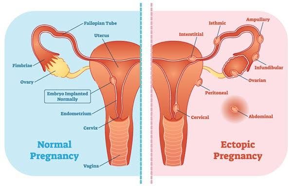
Ectopic pregnancy represents a significant and potentially life-threatening complication of early gestation. Unlike a typical pregnancy where the fertilized egg implants and develops within the uterus, an ectopic pregnancy occurs when implantation takes place outside the uterine cavity. This abnormal location prevents viability and poses serious risks to maternal health. Understanding the nuances of this condition, from its definition and presentation to its diagnosis and management, is crucial for timely intervention and improved outcomes.
Definition of Ectopic Pregnancy
An ectopic pregnancy is defined as the implantation of a fertilized ovum in any location other than the endometrial lining of the uterine cavity. The term “ectopic” originates from the Greek word “ektopos,” meaning “out of place.”
The vast majority of ectopic pregnancies, approximately 95-98%, occur in the fallopian tube. These are often referred to as tubal pregnancies. Within the fallopian tube, implantation can occur in different segments:
- Ampulla (most common): The wider, outer portion of the tube.
- Isthmus: The narrow portion connecting to the uterus.
- Fimbria: The finger-like projections at the end of the tube near the ovary.
- Interstitial or Cornual: The portion of the tube that passes through the muscular wall of the uterus (less common but higher risk of severe hemorrhage if ruptured).
While tubal pregnancies are the most prevalent, ectopic implantation can also occur in other extrauterine locations, including:
- Ovary (ovarian pregnancy): Implantation on the ovary surface or within its tissue.
- Abdomen (abdominal pregnancy): Implantation on abdominal structures such as the bowel, mesentery, or diaphragm. This is rare but carries high risks.
- Cervix (cervical pregnancy): Implantation within the cervical canal. This is also rare and associated with a high risk of severe bleeding.
- Cesarean scar (cesarean scar pregnancy): Implantation within the fibrous tissue of a previous cesarean section scar on the uterus. This is increasingly recognized and presents unique management challenges.
Regardless of the specific location, an ectopic pregnancy cannot progress to a viable term pregnancy because the extrauterine tissues are incapable of supporting the necessary growth and development. Furthermore, as the pregnancy grows, it can erode blood vessels and potentially rupture the structure in which it is implanted, leading to internal hemorrhage.
Clinical Presentation of Ectopic Pregnancy
The clinical presentation of ectopic pregnancy is notoriously variable, ranging from subtle and non-specific symptoms to acute, life-threatening emergencies. Healthcare professionals must maintain a high index of suspicion for ectopic pregnancy in any woman of childbearing age presenting with potential early pregnancy symptoms.
The classic triad of symptoms associated with ectopic pregnancy includes:
- Amenorrhea: A missed menstrual period or a positive pregnancy test. This often precedes the onset of other symptoms.
- Abdominal Pain: This is the most common symptom. The pain is typically located in the lower abdomen or pelvis. It can be unilateral (on one side), bilateral, or diffuse. The character of the pain varies; it may be described as cramping, dull, aching, or sharp. The onset can be sudden or gradual. Pain may worsen with movement or defecation.
- Vaginal Bleeding: This usually presents as irregular spotting or light bleeding, often described as brown or dark red. It is typically less heavy than a normal menstrual period or a miscarriage. However, bleeding amount and pattern can vary significantly.
It is crucial to note that this classic triad is not always present. Many patients may have only one or two of these symptoms, or they may present with entirely different manifestations. Other potential symptoms include:
- Shoulder Pain: Often described as pain under the scapula (shoulder blade). This is a referred pain caused by irritation of the diaphragm by blood accumulating in the abdominal cavity (hemoperitoneum), typically after rupture.
- Dizziness or Syncope: Feeling lightheaded, faint, or actually losing consciousness. These symptoms are indicative of significant blood loss and impending or actual hemodynamic instability.
- Rectal Pressure or Pain with Defecation: Can occur due to pelvic irritation from blood or the ectopic mass.
- Gastrointestinal Symptoms: Nausea, vomiting, or diarrhea may occur.
Physical examination findings can also be variable. They may include:
- Lower abdominal tenderness, which may be localized or diffuse.
- Adnexal tenderness or a palpable adnexal mass (though a mass is not always detectable).
- Cervical motion tenderness (pain upon moving the cervix during a pelvic exam).
- Signs of hypovolemic shock if rupture and significant bleeding have occurred (tachycardia, hypotension, pallor, cool extremities).
Given the wide spectrum of presentation, a high level of clinical suspicion is paramount, especially in women with risk factors for ectopic pregnancy (such as a history of pelvic inflammatory disease, prior ectopic pregnancy, tubal surgery, smoking, or use of assisted reproductive technologies).
Risk of Ruptured Ectopic Pregnancy
The primary and most severe risk associated with an ectopic pregnancy is rupture. As the pregnancy grows within the confined space of the fallopian tube or other ectopic site, it stretches and ultimately erodes through the surrounding tissue and blood vessels. This rupture leads to significant internal hemorrhage (bleeding into the abdominal cavity).
A ruptured ectopic pregnancy is a medical emergency requiring immediate surgical intervention. The consequences of rupture and subsequent internal bleeding can be catastrophic:
- Severe Hemorrhage: The rich blood supply in the pelvic region means that rupture can cause rapid and massive blood loss.
- Hypovolemic Shock: Significant blood loss leads to a dramatic drop in blood pressure, inadequate organ perfusion, and symptoms like rapid heart rate, dizziness, confusion, and loss of consciousness.
- Organ Damage: Prolonged shock can lead to damage of vital organs due to lack of oxygen.
- Maternal Mortality: Ectopic pregnancy is a leading cause of maternal death in the first trimester. While mortality rates have significantly decreased with improved diagnosis and management, rupture remains the primary cause of death.
The risk of rupture is influenced by the location of the ectopic pregnancy (interstitial pregnancies have a higher risk of severe hemorrhage upon rupture) and the stage of gestation. Rupture typically occurs within the first 6-12 weeks of pregnancy.
Recognizing the signs of rupture (sudden, severe pain; signs of shock) and acting swiftly is critical for reducing morbidity and mortality. Even without frank rupture, bleeding around the ectopic site can occur, causing pain and potentially leading to a “leaking” ectopic pregnancy. Therefore, any suspected ectopic pregnancy should be evaluated and managed promptly before rupture occurs.
Diagnostic Approach for Suspected Ectopic Pregnancy
The diagnostic evaluation for a patient with suspected ectopic pregnancy aims to confirm the presence of an early pregnancy and determine its location. The approach typically involves a combination of clinical assessment, laboratory tests, and imaging studies.
- Clinical Assessment:
- Detailed History: Gather information about the patient’s menstrual history, sexual activity, contraceptive use, current symptoms (onset, character, location of pain; amount and nature of bleeding), presence of pregnancy symptoms, and risk factors for ectopic pregnancy.
- Physical Examination: Assess vital signs for stability (heart rate, blood pressure). Perform an abdominal and pelvic examination to assess for tenderness, guarding, masses, or signs of peritoneal irritation or shock.
- Laboratory Tests:
- Quantitative Serum Beta-hCG (Human Chorionic Gonadotropin): This test confirms pregnancy and measures the exact level of the pregnancy hormone. In a normal early intrauterine pregnancy, serum hCG levels typically double approximately every 48 hours. In an ectopic pregnancy, the rise in hCG is often slower, plateaus, or may even fall, but this pattern is not definitive and can overlap with non-viable intrauterine pregnancies (miscarriages). The absolute hCG level is used in conjunction with ultrasound findings.
- Progesterone Levels: Serum progesterone levels may also provide supportive information, but they are not diagnostic. Low progesterone levels (< 5 ng/mL) are often associated with non-viable pregnancies (both intrauterine and ectopic), while higher levels are more reassuring for a potentially viable pregnancy, but do not rule out a rare viable heterotopic pregnancy (simultaneous intrauterine and ectopic).
- Complete Blood Count (CBC): To assess for anemia due to bleeding and check platelet count.
- Blood Type and Rh Status: Essential for potential transfusion needs and administration of Rh immunoglobulin (Rhogam) if the patient is Rh-negative.
- Imaging Studies:
- Transvaginal Ultrasound (TVUS): This is the cornerstone of diagnosing or excluding ectopic pregnancy in the hemodynamically stable patient. TVUS provides detailed images of the uterus, ovaries, and adnexa. Key findings include:
- Absence of an Intrauterine Gestational Sac: When the serum hCG level reaches a certain threshold (known as the “discriminatory zone,” typically 1500-2000 mIU/mL), an intrauterine gestational sac should be visible on TVUS in a normal pregnancy. The absence of an intrauterine sac at or above this hCG level is highly suggestive of an ectopic pregnancy or a completed miscarriage.
- Visualization of an Adnexal Mass: An abnormal mass separate from the ovary in the fallopian tube region. This mass may appear as a complex cystic or solid structure and may or may not contain a yolk sac, embryo, or visible cardiac activity.
- Free Fluid in the Cul-de-sac or Abdomen: Echogenic free fluid suggests the presence of blood, indicating hemorrhage, which can be associated with rupture or bleeding from an ectopic pregnancy.
- Transvaginal Ultrasound (TVUS): This is the cornerstone of diagnosing or excluding ectopic pregnancy in the hemodynamically stable patient. TVUS provides detailed images of the uterus, ovaries, and adnexa. Key findings include:
- Serial Evaluation: If the initial hCG is below the discriminatory zone and TVUS is inconclusive (no intrauterine pregnancy or definitive adnexal mass seen), the patient is often managed with serial hCG measurements and repeat TVUS every 48-72 hours. The pattern of hCG rise and the findings on subsequent ultrasounds help differentiate between a normal intrauterine pregnancy, a non-viable intrauterine pregnancy, and an evolving ectopic pregnancy.
- Other Diagnostic Modalities (Less Common):
- Culdocentesis: A procedure to aspirate fluid from the rectouterine pouch (cul-de-sac). The presence of non-clotting blood strongly suggests hemoperitoneum, often due to a ruptured ectopic pregnancy. This is less commonly performed now with the widespread availability of ultrasound.
- Diagnostic Laparoscopy: In uncertain cases, particularly if suspicion remains high despite inconclusive non-invasive tests, or if the patient is hemodynamically borderline, direct visualization via laparoscopy may be necessary to confirm the diagnosis and allow for immediate treatment.
The diagnostic process is often one of exclusion. The goal is to definitively locate the pregnancy. If an intrauterine pregnancy is clearly visualized on ultrasound, ectopic pregnancy is largely ruled out (except for the rare possibility of a heterotopic pregnancy). If an intrauterine pregnancy is not seen and the clinical picture and serial testing are consistent, ectopic pregnancy is the leading diagnosis until proven otherwise.
Principles of Medical and Surgical Management of Ectopic Pregnancy
Once diagnosed, the management of an ectopic pregnancy depends on several factors, including the patient’s clinical stability, the size and characteristics of the ectopic mass, the presence or absence of fetal cardiac activity, the initial hCG level, the patient’s desire for future fertility, and her ability to comply with follow-up. The primary goals of management are to resolve the ectopic pregnancy, prevent rupture, minimize morbidity, and preserve future reproductive potential whenever possible.
Management options are broadly categorized as medical or surgical. Expectant management (close observation without intervention) is sometimes considered for very early, asymptomatic ectopics with falling hCG levels, but this approach requires strict criteria and vigilant monitoring.
A. Medical Management
Medical management is typically used for unruptured ectopic pregnancies under specific conditions. The most common medical treatment involves the use of Methotrexate.
- Methotrexate: A chemotherapy agent that inhibits cell division and growth by interfering with folic acid metabolism. It effectively stops the growth of the ectopic pregnancy tissue.
- Criteria for Medical Management:
- Hemodynamically stable patient.
- No signs of rupture or significant active bleeding.
- Ability and willingness to comply with necessary follow-up.
- Generally, ectopic mass size less than 3-4 cm.
- Absence of fetal cardiac activity on ultrasound.
- Initial serum hCG level often below a certain threshold (e.g., < 5000 mIU/mL, though criteria vary).
- No contraindications (e.g., liver or kidney disease, pre-existing blood disorders, active infection, immunodeficiency, breastfeeding, known sensitivity to methotrexate).
- Administration: Methotrexate is usually given as a single intramuscular injection, though multi-dose regimens exist.
- Follow-up: Close monitoring is essential. Serum hCG levels are measured on specific days after injection (commonly day 4 and day 7) to assess treatment effectiveness. A decrease of at least 15% between day 4 and day 7 is considered a favorable response. Follow-up hCG levels are then checked weekly until they are undetectable (indicating complete resolution).
- Potential Side Effects: Nausea, vomiting, stomatitis (mouth sores), abdominal pain (often occurring a few days after injection due to tissue necrosis), fatigue.
- Failure Rate: Medical management is successful in approximately 70-95% of carefully selected cases. Failure requires surgical intervention.
- Criteria for Medical Management:
B. Surgical Management
Surgical intervention is indicated in several scenarios, including:
- Hemodynamically unstable patient with suspected or confirmed ectopic pregnancy.
- Signs or strong suspicion of ectopic rupture.
- Patient presenting with severe pain.
- Presence of fetal cardiac activity in the ectopic mass.
- Ectopic mass size larger than criteria for medical management.
- Contraindications to methotrexate or inability to comply with medical follow-up.
- Failed medical management (persistently high or rising hCG after methotrexate).
- Patient preference after counseling.
Surgical approaches include:
- Laparoscopy: This minimally invasive surgical technique is the preferred approach for most stable patients requiring surgery. It involves small incisions in the abdomen through which a camera and surgical instruments are inserted. Laparoscopy offers advantages such as smaller scars, less pain, shorter hospital stay, and faster recovery compared to open surgery.
- Laparotomy: Open abdominal surgery is typically reserved for hemodynamically unstable patients with significant intra-abdominal hemorrhage or in situations where laparoscopy is not feasible (e.g., extensive adhesions, very large ectopic masses).
Surgical procedures performed to remove the ectopic pregnancy include:
- Salpingostomy: An incision is made along the length of the fallopian tube over the ectopic pregnancy, and the pregnancy tissue is gently removed, leaving the tube intact. This procedure may be considered for small, unruptured tubal ectopics, particularly in women who desire future fertility and whose contralateral tube is damaged or absent. A risk of persistent ectopic tissue (requiring further treatment) exists with salpingostomy if not all tissue is removed.
- Salpingectomy: The entire fallopian tube containing the ectopic pregnancy is removed. This is the more common procedure, especially if the tube is significantly damaged by the ectopic or rupture, or if the patient has completed childbearing. Salpingectomy effectively eliminates the risk of persistent ectopic tissue in that tube and may reduce the risk of recurrence in the same tube.
C. Post-Treatment Care:
Regardless of the management method, follow-up care is important.
- Rh Status: Rh-negative women diagnosed with ectopic pregnancy require administration of Rh immunoglobulin (Rhogam) to prevent Rh sensitization.
- hCG Monitoring: After both medical and surgical treatment (especially salpingostomy), serial hCG levels are monitored until they are undetectable to ensure complete resolution of the pregnancy tissue.
- Counseling: Patients should receive counseling regarding the experience, emotional support, future pregnancy risks (a history of ectopic pregnancy increases the risk of recurrence), and contraceptive options.
In conclusion, ectopic pregnancy is a critical condition in early pregnancy that demands prompt recognition, accurate diagnosis, and appropriate management based on individual patient factors. A high index of suspicion, timely access to diagnostic tools like hCG testing and transvaginal ultrasound, and a clear understanding of medical and surgical treatment principles are essential for ensuring optimal outcomes and minimizing the significant risks associated with this condition.


