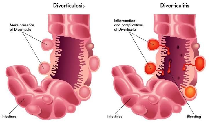
1. Diverticular Disease
Diverticular disease refers to the presence of diverticula – small, bulging pouches that can form in the lining of the digestive tract, most commonly in the lower part of the large intestine (colon). The presence of these pouches is termed diverticulosis. When these pouches become inflamed or infected, the condition is known as diverticulitis.
1.1 Clinical Findings: Differentiating Diverticulosis and Diverticulitis
Understanding the distinction in clinical presentation is crucial for appropriate diagnosis and management.
- Diverticulosis:
- Symptoms: The vast majority of individuals with diverticulosis are completely asymptomatic. The presence of diverticula is often discovered incidentally during investigations for other conditions (e.g., colonoscopy, CT scan). Some individuals may report non-specific symptoms such as mild cramping, bloating, or constipation, often attributed to the underlying bowel motility pattern rather than the diverticula themselves.
- Signs: Physical examination in diverticulosis is typically normal. There are no specific physical signs associated solely with the presence of uncomplicated diverticula.
- Diverticulitis:
- Symptoms: This is a symptomatic condition characterized by inflammation or infection of one or more diverticula.
- Abdominal Pain: This is the hallmark symptom. Pain is typically located in the left lower quadrant (LLQ), as diverticula most commonly affect the sigmoid colon. The pain is often described as constant and cramping, and may be severe. It usually develops gradually over a few hours or days.
- Fever: Elevated temperature is a common sign of inflammation or infection.
- Change in Bowel Habits: This can vary, including constipation, diarrhea, or a sense of incomplete evacuation.
- Nausea and Vomiting: May occur, especially with more severe inflammation or associated partial obstruction.
- Urinary Symptoms: If the inflamed segment of colon is adjacent to the bladder, irritation can lead to urinary urgency, frequency, or dysuria.
- Signs: Physical examination reveals specific findings indicative of peritoneal irritation.
- Localized Tenderness: Significant tenderness upon palpation is present in the affected area, most commonly the LLQ.
- Guarding and Rebound Tenderness: Muscle guarding and rebound tenderness may be present, indicating peritoneal involvement. This suggests more severe inflammation or potential perforation.
- Palpable Mass: In some cases, an inflammatory mass or abscess can be palpable in the LLQ.
- Abdominal Distension: May be present with associated ileus or obstruction.
- Tachycardia: Reflecting pain, fever, or systemic inflammation.
- Symptoms: This is a symptomatic condition characterized by inflammation or infection of one or more diverticula.
1.2 Complications of Diverticular Disease
Complications typically arise from acute diverticulitis and range in severity.
- Abscess Formation: A localized collection of pus forms next to the inflamed diverticulum. This is a common complication (up to 15% of acute uncomplicated diverticulitis cases). Patients present with worsening pain, fever, and often a palpable mass.
- Treatment: Small abscesses (<2-3 cm) may be managed with antibiotics. Larger abscesses typically require percutaneous drainage under CT or ultrasound guidance, in addition to antibiotics. Surgical drainage may be necessary if percutaneous access is not feasible or fails.
- Fistula Formation: Abnormal tracts can form between the inflamed segment of colon and adjacent organs.
- Common types: Colovesical (colon to bladder – leading to pneumaturia, fecaluria, recurrent UTIs), colovaginal (colon to vagina – leading to passage of gas/stool from the vagina), coloenteric (colon to small intestine), colocutaneous (colon to skin).
- Treatment: Fistulas typically require surgical management, usually involving resection of the diseased colonic segment and repair of the affected organ.
- Perforation and Peritonitis: The inflamed diverticulum can rupture, spilling intestinal contents into the abdominal cavity. This can be a localized perforation contained by surrounding structures (leading to abscess) or a free perforation into the peritoneal cavity (leading to diffuse peritonitis).
- Presentation: Sudden onset of severe, diffuse abdominal pain, rigid abdomen, signs of sepsis or shock (tachycardia, hypotension).
- Treatment: Free perforation leading to diffuse peritonitis is a surgical emergency. Treatment involves immediate surgical exploration, lavage of the peritoneal cavity, and typically resection of the perforated segment (often with a temporary stoma, though primary anastomosis may be considered in selected stable patients).
- Obstruction: Chronic inflammation and scarring can lead to narrowing of the colonic lumen, causing partial or complete bowel obstruction. Acute inflammation can also cause temporary obstruction due to associated edema and ileus.
- Presentation: Abdominal distension, colicky abdominal pain, nausea, vomiting, constipation, absence of flatus.
- Treatment: Acute obstructive symptoms due to inflammation often improve with medical management (bowel rest, IV fluids, antibiotics). Chronic strictures causing significant obstruction typically require surgical resection of the narrowed segment.
- Bleeding: While often discussed separately (see Section 3), diverticula are a major cause of acute lower GI bleeding. This is not inflammatory; bleeding occurs when an artery overlying the dome of a diverticulum erodes. It can be minor or massive.
- Treatment: See Section 3.
1.3 Treatment of Diverticular Disease
- Asymptomatic Diverticulosis: No specific treatment is required. A high-fiber diet and adequate hydration are often recommended, though evidence supporting their role in preventing complications is mixed.
- Acute Uncomplicated Diverticulitis:
- Outpatient Management: For mild cases without signs of systemic illness or complications. Bowel rest (clear liquids or low-residue diet), oral antibiotics covering common enteric flora (e.g., ciprofloxacin + metronidazole, or amoxicillin-clavulanate), and pain control.
- Inpatient Management: For more severe pain, fever, signs of systemic illness, inability to tolerate oral intake, or presence of comorbidities. IV fluids, IV antibiotics, bowel rest (NPO initially), pain control. Patients are monitored for clinical improvement or signs of complications.
- Acute Complicated Diverticulitis: Management depends on the specific complication (as detailed above) and typically involves a combination of antibiotics, drainage procedures (percutaneous or surgical), and potentially emergent or elective surgery (resection).
2. Mesenteric Ischemia
Mesenteric ischemia is a condition characterized by insufficient blood flow to the small or large intestine, leading to tissue injury or death (infarction). It can be acute (sudden onset, often severe) or chronic (gradual onset, less severe symptoms). This section focuses primarily on acute mesenteric ischemia due to its critical presentation.
2.1 Clinical Findings and Presentation
Acute mesenteric ischemia is a vascular emergency. Its presentation is often insidious in its early stages but progresses rapidly.
- “Pain Out of Proportion to Physical Findings”: This is the classic hallmark symptom. Patients report severe, diffuse abdominal pain, often sharp or cramping, but the physical examination initially reveals minimal or no abdominal tenderness, guarding, or rigidity. This discrepancy is a critical warning sign.
- Early Symptoms: Nausea, vomiting, diarrhea, or urgent need to defecate are common in the early stages.
- Later Symptoms: As bowel infarction develops, symptoms evolve:
- Abdominal tenderness becomes present and may progress to guarding and rebound as peritonitis sets in.
- Abdominal distension develops.
- Systemic signs of shock or sepsis: Tachycardia, hypotension, fever, altered mental status.
- Bloody stool may occur late as the mucosa sloughs.
- Risk Factors: Patients often have underlying conditions predisposing to vascular compromise:
- Atrial fibrillation or other cardiac arrhythmias (source of emboli).
- Recent myocardial infarction.
- Valvular heart disease.
- Peripheral vascular disease.
- Hypercoagulable states.
- Low cardiac output states (e.g., heart failure, shock – non-occlusive mesenteric ischemia).
2.2 Diagnosis
Prompt diagnosis is vital as prognosis worsens significantly with delaying treatment.
- Clinical Suspicion: High index of suspicion based on severe abdominal pain in a patient with vascular risk factors, especially with the disproportionate pain finding.
- Laboratory Findings: Non-specific initially. May show elevated white blood cell count, metabolic acidosis (lactic acidosis as ischemia worsens), elevated amylase or phosphate (late findings).
- Imaging:
- CT Angiography (CTA): This is the preferred initial diagnostic test. It can visualize the mesenteric vessels (superior and inferior mesenteric arteries and veins) to identify occlusions (embolus, thrombus) or stenosis. It can also show signs of bowel ischemia (bowel wall thickening, pneumatosis intestinalis – air in the bowel wall, portomesenteric venous gas).
- Conventional Angiography: Can be used diagnostically if CTA is equivocal, or therapeutically for intervention (infusion of thrombolytics or vasodilators, angioplasty, stenting). More invasive than CTA.
- Plain X-rays: Usually not helpful in early ischemia, may show non-specific findings like ileus. Late findings like pneumatosis or portal venous gas are poor prognostic signs.
2.3 Treatment
Treatment focuses on restoring blood flow to the bowel as quickly as possible and supporting the patient.
- Resuscitation: Stabilize the patient with IV fluids, manage hypotension, and correct acidosis.
- Pain Control: Provide adequate analgesia.
- Anticoagulation: IV heparin is usually initiated promptly to prevent further clot formation.
- Revascularization: The definitive treatment for occlusive ischemia (embolic or thrombotic).
- Endovascular Therapy: Catheter-directed thrombolysis, angioplasty, stenting. Less invasive than surgery, preferred in stable patients with suitable anatomy.
- Surgical Revascularization: Open embolectomy or bypass surgery. Required for patients who are unstable, have failed endovascular attempts, or whose anatomy is not suitable for endovascular approach.
- Surgical Resection: If bowel infarction has occurred, non-viable segments of bowel must be surgically removed. A “second look” operation within 24 hours may be necessary to assess bowel viability after revascularization.
- Medical Management: For non-occlusive mesenteric ischemia (NOMI), treatment involves optimizing cardiac output and systemic blood pressure and potentially intra-arterial infusion of vasodilators (e.g., papaverine) via angiography.
3. Massive Lower GI Bleeding
Massive lower gastrointestinal (LGI) bleeding is defined as severe bleeding distal to the ligament of Treitz, resulting in hemodynamic instability (persistent hypotension, tachycardia, need for blood transfusion). It represents a significant clinical challenge.
3.1 Differential Diagnosis
Identifying the source is critical after achieving hemodynamic stability. The most common causes include:
- Diverticular Bleeding: The most frequent cause of acute, massive LGI bleeding (up to 50%). Bleeding occurs from a rupture of an arteriole within the wall of a diverticulum. Characteristically painless and often stops spontaneously (70-90%).
- Angiodysplasia (Arteriovenous Malformation): Dilated, tortuous submucosal vessels, most common in the cecum and right colon. Can cause intermittent or massive bleeding. More common in elderly patients and those with chronic kidney disease or aortic stenosis. Bleeding is typically painless.
- Post-Polypectomy Bleeding: Bleeding can occur immediately after polypectomy or days later (delayed post-polypectomy hemorrhage). Risk depends on polyp size and technique.
- Inflammatory Bowel Disease (IBD): Severe colitis (Ulcerative Colitis more commonly than Crohn’s Disease) can cause significant bleeding due to mucosal ulceration. Usually associated with other symptoms of IBD (diarrhea, abdominal pain, fever).
- Ischemic Colitis: Bleeding is usually mild to moderate, associated with abdominal pain (often transient), and diarrhea. Severe, massive bleeding is less common but can occur in rare cases of transmural ischemia.
- Malignancy: Colorectal cancer can cause chronic occult bleeding or intermittent, small volume bleeding. Massive bleeding is uncommon but possible, typically from ulceration or fistula formation.
- Radiation Proctitis: Inflammation of the rectum following pelvic radiation therapy, causing telangiectasias and mucosal friability that can lead to chronic or acute bleeding.
- Rectal Ulcers: Including solitary rectal ulcer syndrome. Can cause bleeding, often associated with tenesmus and mucus discharge.
- Hemorrhoids and Anal Fissures: Very common causes of bright red blood per rectum, but typically small volume and not massive enough to cause hemodynamic instability.
3.2 Initial Management
Management of massive LGI bleeding is primarily focused on resuscitation and stabilization before attempting diagnosis and definitive treatment.
- Resuscitation:
- Airway, Breathing, Circulation (ABCs): Ensure airway patency and adequate ventilation.
- Vascular Access: Establish at least two large-bore intravenous lines (16-gauge or larger).
- Fluid Resuscitation: Rapid infusion of crystalloid solutions (e.g., normal saline or lactated Ringer’s) to restore intravascular volume.
- Blood Transfusion: Administer packed red blood cells (PRBCs) promptly for hemodynamic instability or significant anemia. Massive transfusion protocols may be initiated in severe cases. Platelets and fresh frozen plasma may be needed to correct coagulopathy or thrombocytopenia, especially with massive transfusion or in patients on anticoagulants/antiplatelet agents.
- Hemodynamic Monitoring: Continuous monitoring of heart rate, blood pressure, and oxygen saturation. Central venous pressure monitoring may be helpful.
- Urinary Catheter: Inserted to monitor urine output as an indicator of renal perfusion.
- Assessment: Rapid history focused on prior bleeding episodes, medication use (aspirin, NSAIDs, anticoagulants, antiplatelets), comorbidities (liver disease, cardiac disease), and previous colonoscopies. Physical exam focusing on vital signs, signs of hypovolemia (cool extremities, delayed capillary refill), abdominal exam, and rectal exam (check for hemorrhoids, fissures, masses, stool color – melena vs. hematochezia).
- Laboratory Tests: Complete blood count (CBC), coagulation profile (PT, PTT, INR, platelets), electrolytes, renal function tests, liver function tests, blood type and crossmatch.
- Correction of Coagulopathy: Reverse any underlying coagulopathy if possible (e.g., Vitamin K or FFP for elevated INR, platelet transfusion for severe thrombocytopenia).
3.3 Appropriate Diagnostic Tests
Once the patient is hemodynamically stable, efforts focus on identifying the source of bleeding.
- Colonoscopy: The preferred initial diagnostic modality after adequate bowel preparation. Can identify and often treat various sources (diverticula, angiodysplasia, post-polypectomy sites, IBD lesions). Bowel preparation in the setting of active bleeding can be challenging but is often necessary for adequate mucosal visualization. Rapid lavage protocols are sometimes used.
- CT Angiography (CTA): Useful if the bleeding is active (>0.3-0.5 mL/min) and the patient is too unstable for bowel prep/colonoscopy or if colonoscopy is negative. Can localize the bleeding source by identifying contrast extravasation into the bowel lumen. Can also identify structural lesions.
- Nuclear Medicine Scan (Tagged Red Blood Cell Scan): Highly sensitive for detecting slower rates of active bleeding (>0.05-0.1 mL/min), even when intermittent. Less precise in localization than CTA or angiography but can indicate the general region of bleeding.
- Conventional Angiography: Can localize actively bleeding vessels and allows for therapeutic intervention (embolization). Often used when bleeding is rapid, CTA suggests a source, or colonoscopy fails to identify or control the bleeding.
3.4 Treatment
Treatment depends on the identified source and severity of bleeding.
- Endoscopic Therapy: First-line treatment for many identifiable sources during colonoscopy:
- Diverticular Bleeding: Clipping, band ligation, thermal coagulation (less common due to risk of perforation), epinephrine injection.
- Angiodysplasia: Argon plasma coagulation (APC) or thermal coagulation.
- Post-Polypectomy Bleeding: Clipping, thermal coagulation, epinephrine injection.
- Angiographic Embolization: Performed during conventional angiography. A catheter is advanced to the bleeding vessel, and embolizing agents (coils, particles) are injected to occlude the vessel and stop bleeding. Highly effective for diverticular and angiodysplastic bleeding when endoscopic methods fail or are not feasible due to active, rapid bleeding.
- Surgery: Reserved for patients with persistent severe bleeding refractory to endoscopic and angiographic interventions, recurrent bleeding, or complications (e.g., perforation).
- If the bleeding source is precisely localized (e.g., by angiography), subtotal colectomy or segmental resection (hemicolectomy) of the affected colon may be performed.
- If the source cannot be definitively localized and bleeding is life-threatening, a subtotal or total colectomy may be necessary as a last resort to remove all potential sources.
This guide provides a focused professional overview of these distinct yet sometimes related gastrointestinal conditions. Mastery of their clinical presentation, potential complications, and appropriate diagnostic and treatment pathways is essential for effective patient care.


