Emergency Room Management of Spinal Trauma
Spinal trauma is a critical and potentially devastating injury that demands rapid, systematic assessment and intervention in the emergency room (ER) setting. Missed diagnoses or delayed management can lead to permanent neurological deficits or even death.
Initial Assessment, Stabilization, and Spinal Precautions
The immediate priority in any trauma patient is the primary survey following Advanced Trauma Life Support (ATLS) principles: Airway, Breathing, and Circulation (ABCs). Concurrently, aggressive spinal precautions must be initiated and maintained from the moment the patient is encountered.
- A (Airway): Assess for airway patency. Consider potential for cervical spine injury influencing airway management, especially in patients with facial trauma, altered mental status, or high suspicion of cervical fracture/dislocation. Manual in-line stabilization must be maintained during airway maneuvers. If intubation is required, use techniques that minimize cervical spine movement (e.g., rapid sequence intubation with manual in-line stabilization, consideration of fiberoptic intubation).
- B (Breathing): Assess respiratory effort, rate, and oxygenation. High cervical cord injuries can compromise diaphragmatic function (C3-C5 innervation), leading to respiratory failure. Ensure adequate oxygenation (aim >95%) and ventilation.
- C (Circulation): Assess pulse, blood pressure, and signs of shock. Consider both hemorrhagic shock from associated injuries and neurogenic shock (hypotension and bradycardia) stemming from autonomic dysfunction in certain spinal cord injuries (typically T6 or higher). Maintain adequate perfusion, especially to the spinal cord.
- D (Disability): Perform a brief neurological assessment including level of consciousness (GCS), pupillary response, and a quick check for gross motor/sensory deficits. Note any paralysis, weakness, or paresthesias.
- E (Exposure/Environment): Fully expose the patient to identify all injuries, but prevent hypothermia, which can worsen outcomes.
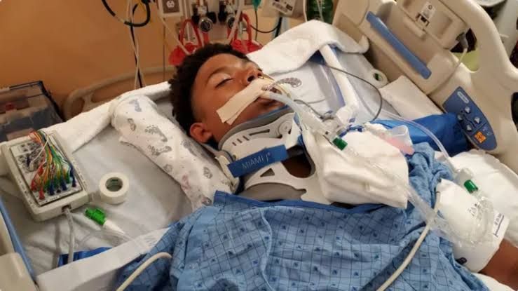
- Spinal Immobilization: All patients with potential spinal trauma based on mechanism (e.g., high-speed motor vehicle collision, fall from height, axial load injury, pedestrian struck) or symptoms (midline spinal pain, neurological deficits, altered mental status, intoxication, distracting injury) must have their spine rigorously immobilized. This involves a rigid cervical collar, placement on a backboard or vacuum mattress (preferably), and secure strapping. Maintain immobilization during all transfers and diagnostic procedures until the spine is cleared.
Detailed History and Physical Examination
Once the patient is stable and immobilized, perform a focused history and a comprehensive physical examination.
- History:
- Mechanism of Injury (MOI): Crucial for determining the likelihood and type of spinal injury (e.g., flexion, extension, compression, distraction, rotation). Key details include height of fall, speed of vehicle impact, use of restraints, position of impact, neurological status reported at the scene.
- Symptoms: Assess presence and nature of spinal pain (location, radiation), numbness, tingling, weakness, paralysis, difficulty breathing, loss of bladder or bowel control. Inquire about pre-existing spine conditions (arthritis, fusion, previous surgery).
- Physical Examination:
- General: Assess vital signs, level of consciousness.
- Spine: Carefully inspect and palpate along the entire vertebral column (cervical, thoracic, lumbar, sacral) for tenderness, deformities, step-offs, gaps between spinous processes, or ecchymosis. Palpation should be performed cautiously to avoid exacerbating injury.
- Neurological Examination: Perform a detailed neurological exam. This includes:
- Motor function: Assess strength in all major muscle groups (e.g., C5-T1 for upper extremities, L2-S1 for lower extremities), ideally using the ASIA (American Spinal Injury Association) motor score scale if feasible, or at minimum documenting ability to move against gravity/resistance.
- Sensory function: Assess light touch and pinprick sensation throughout dermatomes. Document sensory levels precisely.
- Reflexes: Deep tendon reflexes (DTRs) and presence/absence of pathological reflexes (Babinski).
- Rectal Exam: Assess perianal sensation and voluntary external anal sphincter contraction (sacral sparing is critical for prognostication in complete injuries).
Decision Making for Imaging
Not every trauma patient requires immediate spine imaging, particularly plain films of the entire spine. Clinical decision rules like the Canadian C-Spine Rule or the NEXUS criteria can help identify patients at low risk for cervical spine injury who may not require imaging, provided they are reliable examiners. However, in high-energy trauma mechanisms, patients with altered mental status, intoxication, significant distracting injuries, neurological deficits, or midline spinal tenderness, imaging is mandatory.
Imaging Modalities and Interpretation
Imaging is essential to diagnose fractures, dislocations, ligamentous injuries, and spinal cord involvement.
- Plain Radiography (X-rays):
- Role: Historically the primary tool, now largely supplanted by CT for clearing the spine in significant trauma due to limited sensitivity. Still useful as a screening tool in low-risk cases or when CT is unavailable.
- Views (C-spine): Lateral, AP, Odontoid (open mouth). A complete lateral view must visualize the C7-T1 junction. swimmer’s view may be needed if C7 is not seen.
- Interpretation: Systematically review the ABCs: Alignment (anterolisthesis/retrolisthesis, lordosis/kyphosis curves – check anterior, posterior, spinolaminar lines), Bone (fractures, height loss, endplate fx, facet joints), Cartilage/Discs (disc space height), Soft tissue (prevertebral space widening suggesting hematoma/ligamentous injury). Lumbar and thoracic films require AP and Lateral views.
- Computed Tomography (CT Scan):
- Role: The modality of choice for evaluating bony anatomy in trauma due to high sensitivity for fractures and dislocations. Essential for visualizing the spinal canal.
- Views: Axial slices are standard, but sagittal and coronal reconstructions are critical for assessing alignment, facet joint integrity, and subtle fractures.
- Interpretation: Review bone windows meticulously in all planes. Look for fracture lines, step-offs, facet joint abnormalities (perched or locked facets), malalignment, and retropulsed bone fragments encroaching on the spinal canal. Compare canal size to expected norms. Perform 3D reconstructions if available for complex fractures.
- Magnetic Resonance Imaging (MRI):
- Role: Essential for assessing soft tissue structures (ligaments, discs), evaluating the spinal cord parenchyma itself, identifying epidural hematoma, or visualizing non-bony causes of compression. Indicated for patients with a neurological deficit, suspected ligamentous injury despite negative/equivocal CT, or unexplained spinal pain after negative plain films/CT.
- Interpretation: Review T1, T2, and STIR sequences. Look for: spinal cord edema (high signal on T2/STIR, suggests contusion), hemorrhage (variable signal, often low on T2*), compression (extrinsic pressure on cord from disc, ligament, bone, hematoma), ligamentous disruption (high signal on T2/STIR).
Recognizing Spinal Cord Injury (SCI)
SCI is suspected based on neurological findings (weakness, paralysis, sensory loss below a specific level).
- Complete vs. Incomplete SCI:
- Complete: Absence of sensory and motor function in the lowest sacral segments (S4/S5). Prognosis for recovery is generally poor below the level of injury. Spinal shock may initially mask true completeness.
- Incomplete: Preservation of some sensory or motor function below the neurological level, including sensory or motor function in the lowest sacral segments (sacral sparing). Patterns include Central Cord Syndrome, Anterior Cord Syndrome, Brown-Séquard Syndrome, Conus Medullaris Syndrome, Cauda Equina Syndrome. Prognosis for recovery is generally better than complete SCI.
- Spinal Shock: A temporary state of depressed spinal reflexes, motor, and sensory function below the level of injury that occurs immediately after SCI. Resolution (marked by return of reflexes, esp. bulbocavernosus) can take hours to days. Absence of reflexes in the acute phase does not definitively mean a complete injury until spinal shock resolves.
- Neurogenic Shock: A distributive form of shock seen in SCI above T6 due to disruption of descending sympathetic pathways. Characterized by hypotension, bradycardia, and peripheral vasodilation (warm, dry skin). This is distinct from spinal shock (neurological deficit) and hypovolemic shock (tachycardia, cool extremities).
Acute Management of Suspected or Diagnosed SCI
Immediate management focuses on preventing secondary injury to the cord.
- Continued Immobilization: Maintain rigorous spinal immobilization until a definitive plan is in place.
- Systemic Measures (Preventing Secondary Injury):
- Maintain Spinal Cord Perfusion: Target a Mean Arterial Pressure (MAP) > 85-90 mmHg, particularly for the first 7 days (guidelines vary). This is critical to perfuse the injured and potentially ischemic cord. Use intravenous fluids cautiously (especially in neurogenic shock where vasodilation is primary issue) and primarily vasopressors (e.g., norepinephrine, phenylephrine) to achieve the MAP target.
- Optimize Oxygenation: Maintain SpO2 >95%. Provide supplemental oxygen. Monitor respiratory status closely, especially with high cervical injuries. Prophylactic intubation may be necessary if vital capacity is compromised or fatigue is anticipated.
- Temperature Control: Prevent hypothermia.
- Fluid Management: Maintain euvolemia. Avoid aggressive fluid resuscitation solely for hypotension in neurogenic shock; focus on pressors after initial fluid bolus.
- Corticosteroids: The use of high-dose methylprednisolone for acute SCI is highly controversial and is generally not recommended based on meta-analyses and current guidelines (including those from the American Association of Neurological Surgeons/Congress of Neurological Surgeons). While older studies (NASCIS trials) suggested potential marginal benefit if given within 8 hours, this was associated with increased complications (sepsis, pneumonia, GI bleed) and the evidence is considered insufficient to warrant routine use. Current practice favors aggressive supportive care and addressing surgical decompression if indicated.
Understanding the Definition and Management Principles of the Unstable Spine
An unstable spine is a spine that has lost its structural integrity to the extent that it is unable to withstand normal physiological loads without the potential for progressive deformity or causing neurological damage. Instability results from significant bony disruption, ligamentous injury, or facet joint compromise.
- Defining Instability: While complex classification systems exist, in the ER, suspicion of instability is raised by:
- Significant fractures or dislocations on imaging.
- Facet joint abnormalities (perched or locked facets).
- Evidence of significant ligamentous injury (e.g., widening of interspinous distance, facet joint widening/perched, disc extrusion on MRI).
- Progressive neurological deficit after initial injury.
- Management Principles:
- Continued Rigid Immobilization: Absolutely paramount to prevent further injury.
- Reduction (if necessary): Certain injuries like facet dislocations require urgent reduction, sometimes performed in the ER under sedation by experienced personnel, or more commonly in the operating room.
- Surgical vs. Non-Surgical: Unstable injuries generally require surgical stabilization to restore alignment, decompress neural elements, and prevent future deformity/neurological decline. Non-surgical management (e.g., bracing) is reserved for stable fracture patterns without significant neurological deficit.
- Urgent Consultation: Immediate consultation with neurosurgery or orthopedic spine specialists is required for definitive management planning.
Indications for Decompressive Surgery in SCI
The decision for surgical decompression is made by surgical specialists but is often considered for:
- Persistent Spinal Cord Compression: Especially in incomplete SCI, if compression by bone (e.g., burst fracture), disc, or hematoma is evident on imaging and potentially reversible. Early decompression may improve neurological recovery, though the optimal timing is still debated (some evidence suggests benefit within 24 hours).
- Irreducible Facet Dislocations: Requires reduction and stabilization, usually surgical.
- Gross Spinal Instability: To prevent further injury and restore biomechanical integrity.
- Progressive Neurological Deficit: While initially stable, a patient showing signs of worsening neurological function may need urgent imaging and consideration for decompression.
Initial Management of Medical Complications Associated with Cord Injury
SCI patients are vulnerable to specific complications requiring proactive management in the ER.
- Respiratory Complications: High cervical injuries (C1-C5) can cause diaphragmatic paralysis and respiratory failure. Lower injuries can impair intercostal muscle function and cough, leading to atelectasis and pneumonia.
- Management: Close respiratory monitoring (pulse oximetry, capnography, blood gases), aggressive pulmonary hygiene (suctioning), chest physiotherapy, assisted coughing, and early consideration for intubation and mechanical ventilation.
- Cardiovascular Complications (Neurogenic Shock): Hypotension and bradycardia due to sympathetic denervation.
- Management: Maintain MAP > 85-90 mmHg using vasopressors (phenylephrine, norepinephrine). Avoid excessive fluids alone. Atropine or pacing may be needed for severe bradycardia.
- Bladder Dysfunction: Acute SCI results in an atonic, distended bladder leading to urinary retention.
- Management: Urgent insertion of a Foley catheter to prevent bladder over-distension and potential damage. Strict input/output monitoring.
- Bowel Dysfunction: A paralytic ileus is common in acute SCI, particularly in thoracic and higher injuries, due to loss of autonomic tone. Loss of voluntary control leads to risk of constipation.
- Management: Nasogastric tube insertion for decompression if ileus is suspected or patient is nauseous/vomiting. Bowel regimen initiated early during admission (once stable).
- Skin Complications: SCI patients are at high risk for pressure ulcers due to immobility and sensory loss.
- Management: Careful handling during transfers. Ensure proper padding, especially over bony prominences, while immobilized on a backboard. While frequent turning isn’t possible on a backboard, minimizing time on the board and ensuring proper padding are key initial steps.
Consultation and Disposition
- Consultation: Immediate consultation with the appropriate surgical service (Neurosurgery or Orthopedic Spine Surgery) is mandatory once spinal trauma is identified or strongly suspected, especially with neurological deficit or instability. Other services (e.g., Urology for complex bladder issues, Pulmonary/ICU for respiratory compromise) may be needed.
- Disposition: Patients with confirmed or highly suspected spinal trauma, especially with SCI or instability, require admission to a facility equipped to manage these injuries, often a trauma center with specialized spine care and rehabilitation services.
Conclusion
The emergency room management of spinal trauma is a challenging but critical process. A systematic, step-by-step approach beginning with immediate stabilization and rigorous immobilization, followed by a thorough clinical and radiographic assessment (emphasizing the value of CT and MRI), and culminating in proactive management of potential complications and early consultation with specialists, is essential for optimizing outcomes and preventing further neurological injury. Continuous vigilance for subtle signs, understanding the implications of imaging findings, and maintaining spinal cord perfusion are cornerstones of acute care.


