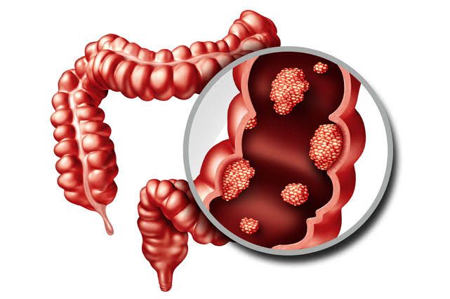
Carcinoma of the colon and rectum, often referred to collectively as colorectal cancer (CRC), is a significant health concern globally. It arises from the inner lining of the large intestine (colon or rectum), typically developing slowly over many years from precancerous polyps. Early detection and timely intervention are crucial for improving outcomes.
Recognizing the Potential Signs and Symptoms
The early stages of colorectal carcinoma often present with no symptoms, which underscores the importance of regular screening. When symptoms do occur, they can be varied and often non-specific, potentially mimicking other less serious conditions. However, persistent changes should prompt medical evaluation. Common signs and symptoms include:
- Changes in Bowel Habits: This is one of the most frequent indicators. It can manifest as persistent diarrhea, constipation, or a change in the consistency of stool that lasts for more than a few weeks. The change might also involve a feeling of incomplete evacuation or urgency.
- Blood in the Stool: This can appear as bright red blood (hematochezia) or dark, tarry stools (melena). Blood may be mixed with the stool, Streak it, or be noticed on toilet paper. While often attributed to hemorrhoids or fissures, any new occurrence of blood in the stool warrants investigation.
- Abdominal Discomfort or Pain: Cramping, bloating, fullness, or general abdominal pain can occur, especially if the tumor is causing a partial or complete blockage of the bowel (obstruction).
- Unexplained Weight Loss: Losing weight without trying can be a symptom of advanced disease.
- Fatigue or Weakness: Cancer can cause iron-deficiency anemia due to chronic, slow bleeding in the digestive tract. Anemia leads to insufficient oxygen transport, resulting in fatigue, weakness, and paleness (pallor).
- A Feeling That the Bowel Doesn’t Empty Completely: This sensation, also known as tenesmus, is more common with rectal tumors.
- A Lump in the Abdomen or Rectum: While less common, a palpable mass may be detected during a physical examination, especially in later stages.
It is critical to remember that these symptoms can be caused by conditions other than cancer. However, their persistence necessitates consultation with a healthcare professional for proper evaluation.
Diagnostic Evaluation
If symptoms suggest the possibility of colorectal carcinoma or if screening tests yield abnormal results, a series of diagnostic studies are performed to confirm the presence of cancer, determine its location, and assess its extent.
- Laboratory Studies:
- Complete Blood Count (CBC): Often ordered to check for anemia, especially iron-deficiency anemia, which can be caused by chronic blood loss from a tumor.
- Liver Function Tests (LFTs): May be performed to assess liver health, as the liver is a common site for colorectal cancer metastasis.
- Carcinoembryonic Antigen (CEA): While not used for initial diagnosis, baseline CEA levels are often measured. CEA is a tumor marker that can be elevated in CRC, but it is not specific to this cancer and can be elevated in other conditions. Its primary role is in monitoring treatment response and detecting recurrence.
- Stool-based tests: While primarily for screening (Fecal Occult Blood Test – FOBT, Fecal Immunochemical Test – FIT, Multi-target stool DNA test), a positive result prompts further diagnostic investigation like colonoscopy.
- Endoscopic Studies:
- Colonoscopy: This is the gold standard for the diagnosis of colorectal carcinoma. A flexible tube with a camera (colonoscope) is inserted into the rectum and advanced through the colon to visualize the entire large intestine. This allows for direct visualization of polyps or tumors. Crucially, biopsies of suspicious lesions can be taken during colonoscopy. A tissue biopsy examined under a microscope by a pathologist is required for a definitive diagnosis of cancer.
- Flexible Sigmoidoscopy: Similar to colonoscopy but examines only the lower part of the colon and the rectum. It can be useful but may miss cancers higher up in the colon.
- Proctoscopy: Examination of the rectum only.
- Imaging Studies (Including X-ray Based): Imaging helps to determine the size and location of the tumor, assess local invasion, and detect potential spread (metastasis) to other organs or lymph nodes.
- Computed Tomography (CT) Scan: Frequently used for staging. A CT scan creates detailed cross-sectional images of the abdomen and pelvis to assess the extent of the primary tumor and identify metastasis in lymph nodes, liver, lungs, or other organs. A chest X-ray or CT chest scan is also often performed to check for lung metastases.
- Magnetic Resonance Imaging (MRI): Particularly valuable for rectal cancer to assess the depth of tumor invasion into the rectal wall and involvement of surrounding structures and lymph nodes. This is crucial for planning treatment, especially radiotherapy and surgery.
- Barium Enema: Less commonly used now, this involves coating the colon lining with barium contrast and taking X-rays. It can reveal polyps or tumors but does not allow for biopsy.
- Positron Emission Tomography (PET) Scan: Often combined with CT (PET-CT). Used in some cases to detect distant metastases or evaluate recurrence, especially when other imaging is inconclusive.
Staging the Disease
Once colorectal carcinoma is diagnosed, staging is performed to determine the extent of the cancer. Staging is critical for guiding treatment decisions and providing a prognosis. Two commonly used staging systems are the TNM system and the modified Dukes classification.
- TNM Classification (AJCC/UICC Staging): This is the most widely used and detailed system. It describes the cancer based on three key components:
- T (Tumor): Describes the size of the primary tumor and how deeply it has invaded the wall of the colon or rectum. T stages range from Tis (carcinoma in situ) or T1 (invading submucosa) up to T4 (invading through the wall and potentially into nearby organs/structures).
- N (Nodes): Indicates whether the cancer has spread to nearby lymph nodes. N0 means no regional lymph node involvement, while N1 and N2 indicate increasing numbers or involvement of regional lymph nodes.
- M (Metastasis): Indicates whether the cancer has spread to distant sites (metastasis). M0 means no distant metastasis, while M1 means distant metastasis is present (e.g., liver, lungs, bone). These T, N, and M values are combined to assign an overall stage (Stage 0, I, II, III, or IV). Higher stages indicate more advanced disease.
- Dukes Classification: An older, simpler system still sometimes referenced, particularly for historical comparison.
- Dukes A: Tumor is limited to the bowel wall.
- Dukes B: Tumor extends through the bowel wall but has not spread to lymph nodes.
- Dukes C: Cancer has spread to regional lymph nodes.
- Dukes D: Cancer has spread to distant organs (metastasis).
5-Year Survival Rate and Staging: The 5-year survival rate is a statistical measure indicating the percentage of people who are still alive five years after diagnosis. It varies significantly depending on the stage at diagnosis:
- Localized (Stage I & II / Dukes A & B): Cancer has not spread significantly beyond the colon or rectum wall. The 5-year survival rate is typically very high (e.g., >90% for Stage I, 70-85% for Stage II).
- Regional (Stage III / Dukes C): Cancer has spread to regional lymph nodes but not distant sites. The 5-year survival rate is lower but still significant (e.g., 50-70%).
- Distant (Stage IV / Dukes D): Cancer has spread to distant organs. The prognosis is significantly poorer, with 5-year survival rates typically in the range of 10-20%, although treatment advances are improving these figures, especially with targeted therapies.
These statistics are averages and do not predict the outcome for any individual patient. Many factors influence prognosis, including overall health, tumor characteristics, and response to treatment.
Treatment Options
Treatment for colorectal carcinoma is multidisciplinary, involving surgeons, oncologists (medical and radiation), pathologists, radiologists, and other specialists. The specific treatment plan depends primarily on the stage of the cancer, its location (colon vs. rectum), the patient’s overall health, and individual preferences.
- Surgery: This is the primary treatment for most stages of colorectal carcinoma that have not widely metastasized. The goal is to remove the section of the colon or rectum containing the tumor, along with a margin of healthy tissue and nearby lymph nodes.
- Colectomy: For colon cancer, surgery involves removing part or all of the colon (e.g., hemicolectomy, sigmoidectomy). The remaining sections of the colon are usually reconnected (anastomosis).
- Proctectomy: For rectal cancer, surgery involves removing part or all of the rectum. Depending on the tumor’s location and extent, it may be possible to reconnect the colon to the remaining rectum or anal canal (low anterior resection – LAR). In some cases, a permanent colostomy (opening of the colon to the outside of the body to divert stool) may be necessary (e.g., abdominoperineal resection – APR).
- Chemotherapy: The use of drugs to kill cancer cells.
- Adjuvant Chemotherapy: Given after surgery, typically for Stage II high-risk or Stage III colon and rectal cancers, to destroy any remaining microscopic cancer cells and reduce the risk of recurrence.
- Neoadjuvant Chemotherapy: Given before surgery, more commonly for rectal cancer, often combined with radiation (radiochemotherapy). Its goals are to shrink the tumor, making surgery easier and potentially allowing for sphincter preservation in rectal cancer, and to reduce local recurrence.
- Radiotherapy (Radiation Therapy): The use of high-energy rays to kill cancer cells. Radiotherapy is a standard part of treatment for many rectal cancers but is less commonly used for colon cancer unless for palliative purposes (like pain relief) or in locally advanced, unresectable cases. It is frequently given in combination with chemotherapy (radiochemotherapy) before surgery for rectal cancer.
- Radiochemotherapy: Combining radiation therapy and chemotherapy. This combination is particularly effective for locally advanced rectal cancer. The chemotherapy drugs can make cancer cells more sensitive to radiation, enhancing its effectiveness.
- Targeted Therapy and Immunotherapy: Newer drugs that target specific molecular pathways in cancer cells or boost the body’s immune system to fight cancer. These are more commonly used for advanced or metastatic disease (Stage IV) and depend on specific molecular characteristics of the tumor (e.g., gene mutations like KRAS, NRAS, BRAF or mismatch repair deficiency).
Postoperative Management and Follow-Up
Following treatment, regular follow-up is essential to monitor recovery, detect any recurrence of cancer at an early, potentially curable stage, and screen for new polyps or cancers. The follow-up schedule and specific tests are tailored to the individual patient based on the stage of their cancer, treatment received, and risk factors.
Typical follow-up includes:
- Clinical Examination: Regular visits with the oncologist and/or surgeon, including a physical exam.
- Laboratory Tests:
- Carcinoembryonic Antigen (CEA): Serum CEA levels are typically checked regularly during follow-up, often every few months for the first few years, then less frequently. While not diagnostic of recurrence on its own, a persistent rise in CEA levels from baseline can be an early indicator of potential recurrence, sometimes occurring before imaging or symptoms. It prompts further investigation.
- Routine blood tests (CBC, LFTs) may also be monitored.
- Imaging Studies:
- CT Scans: Usually performed periodically (e.g., annually for several years) to check for recurrence in the abdomen, pelvis, and chest.
- Endoscopic Surveillance:
- Colonoscopy: Performed at regular intervals (e.g., 1 year after surgery, then every 3-5 years) to look for recurrent tumors at the surgical site, detect new polyps that could develop into cancer, and identify new primary colorectal cancers.
The intensity and duration of follow-up depend on the initial stage and risk of recurrence. Follow-up schedules are typically more frequent in the first 2-3 years after treatment, as this is when most recurrences occur, and may continue for 5 years or longer.
The Role of CEA in Detecting Recurrence:
CEA is a glycoprotein produced by colorectal cancer cells and some normal cells. As mentioned earlier, it’s not specific for CRC and can’t be used for diagnosis, nor is it elevated in all CRC cases. However, if a patient has elevated CEA levels before treatment that fall after successful surgery, or if it was within the normal range pre-treatment but begins to rise during follow-up, it can serve as a valuable marker for monitoring. A rising CEA level during follow-up should trigger further investigation with imaging (like CT scans) to look for the source of the increase. It’s important to interpret CEA results in the context of the patient’s overall clinical picture and other test results. Not all recurrences cause a rise in CEA, and a rise in CEA can sometimes be due to non-cancerous conditions (e.g., smoking, inflammation).
Conclusion
Understanding the signs and symptoms, undergoing appropriate timely diagnostic tests, accurately staging the disease, and receiving comprehensive treatment are all critical steps in managing colorectal carcinoma. Post-treatment follow-up, including clinical evaluation, imaging, endoscopy, and monitoring tumor markers like CEA, plays a vital role in detecting recurrence early, improving the chances of successful salvage therapy. A multidisciplinary approach throughout the patient’s journey offers the best possible outcomes. Early detection through screening remains the most effective strategy for reducing the incidence and mortality of this disease.


