The thyroid gland, a small endocrine organ located at the base of the neck, plays a critical role in regulating metabolism through the production of thyroid hormones (primarily thyroxine, T4, and triiodothyronine, T3). Disorders of the thyroid are common and can significantly impact overall health and well-being. Accurate diagnosis and appropriate treatment are essential for managing these conditions.
Understanding the Thyroid Isotope Scan: Technique and Principles
The thyroid isotope scan, also known as a thyroid scintigraphy or radionuclide scan, is a nuclear medicine imaging test that evaluates the function and structure of the thyroid gland. It relies on the principle that thyroid cells actively take up iodine, a necessary component for hormone synthesis. Radioactive isotopes of iodine are used as tracers to visualize this uptake process.
Principles:
- Thyroid follicular cells have a unique ability to actively transport iodine from the bloodstream into the cell via the sodium-iodide symporter (NIS).
- Once inside the cell, iodine is used to synthesize thyroid hormones.
- Radioactive isotopes of iodine (commonly Iodine-123, I-123) or other tracers that mimic iodine uptake (like Technetium-99m pertechnetate, Tc-99m) are administered to the patient.
- These tracers are taken up by the thyroid cells and emit gamma rays.
- A specialized camera (gamma camera) detects these gamma rays to create images showing the distribution and intensity of tracer uptake within the thyroid gland.
Technique: A Step-by-Step Guide
The process of undergoing a thyroid isotope scan typically involves several steps:
- Step 1: Preparation
- Patient Instructions: The patient receives specific instructions well in advance of the scan. This often includes fasting for several hours (typically 4-6 hours) before tracer administration to optimize intestinal absorption.
- Medication Review: The physician or nuclear medicine staff will review the patient’s medication history. Certain medications, particularly those containing iodine (like amiodarone) or thyroid medications (thyroid hormone replacement, anti-thyroid drugs), can interfere with tracer uptake and may need to be stopped for a specified period before the scan.
- Contrast Media: Recent exposure to iodine-containing contrast media used in CT scans or angiograms can also block thyroid uptake and requires a delay before the scan can be performed (often several weeks).
- Pregnancy and Breastfeeding: Women are routinely screened for pregnancy, as radioactive tracers are contraindicated. Breastfeeding mothers will receive specific instructions regarding pumping and discarding milk after the scan.
- Step 2: Tracer Administration
- The radioactive tracer (most commonly I-123 in capsule or liquid form, or Tc-99m pertechnetate administered intravenously) is given to the patient. The choice of tracer depends on the specific information required and local availability. I-123 is often preferred for evaluating iodine uptake and persistence, while Tc-99m provides images more quickly but only reflects initial uptake, not organification.
- The dose of the tracer is very small and measured in microcuries (µCi) or megabecquerels (MBq).
- Step 3: Waiting Period (Uptake Time)
- Following tracer administration, there is a waiting period to allow the tracer to be absorbed into the bloodstream and subsequently taken up by the thyroid gland.
- The duration of this waiting period depends on the tracer used:
- For Tc-99m, imaging is typically done 20-30 minutes after injection.
- For I-123, imaging is usually performed 4-6 hours and often again at 24 hours after administration to assess both early and delayed uptake and retention.
- Step 4: Imaging Procedure
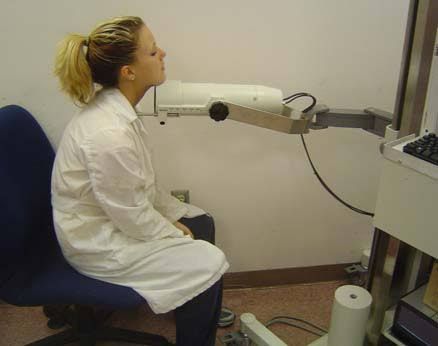
- The patient is positioned on a table, and a gamma camera is placed close to the neck.
- Images of the thyroid gland are acquired from different angles (usually anterior, and often oblique views).
- The patient is required to remain still during the imaging process, which typically takes between 15-30 minutes.
- In addition to the scan images showing the tracer distribution, an uptake measurement is often performed, especially when evaluating hyperthyroidism. This involves measuring the percentage of the administered dose that is taken up by the thyroid gland at specific time points (e.g., 4 and/or 24 hours for I-123). This quantitative measurement is crucial in determining the cause of thyrotoxicosis and planning radio-iodine therapy dosages.
- Step 5: Image Interpretation and Reporting
- A nuclear medicine physician interprets the images and the uptake measurements.
- They evaluate the size, shape, position, and pattern of tracer distribution within the thyroid gland.
- Findings might include:
- Diffuse uniform uptake (consistent with a normal thyroid or Graves’ disease).
- Heterogeneous or patchy uptake (could be multinodular goiter).
- Areas of increased uptake (“hot” nodules or areas).
- Areas of decreased or absent uptake (“cold” nodules or areas).
- Overall low uptake (suggestive of thyroiditis or exogenous hormone intake).
- A formal report is generated summarizing the findings and their clinical significance.
Safety and Side Effects: The dose of radiation from a diagnostic thyroid scan is low and comparable to other standard diagnostic imagine procedures like a CT scan. Side effects are rare but can include mild nausea or allergic reactions to the tracer (very unusual). The benefits of accurate diagnosis typically outweigh these minimal risks.
The Concept of Thyrotoxicosis
Thyrotoxicosis is a clinical and biochemical syndrome resulting from excessive levels of thyroid hormones (T4 and/or T3) circulating in the bloodstream. It is important to distinguish thyrotoxicosis from hyperthyroidism. Hyperthyroidism specifically refers to thyrotoxicosis caused by excessive synthesis and secretion of thyroid hormones by the thyroid gland itself (e.g., in Graves’ disease, toxic nodules). Thyrotoxicosis can also be caused by the release of stored hormone (thyroiditis), ingestion of excess hormone (thyrotoxicosis factitia), or production outside the thyroid (very rare).
Causes of Thyrotoxicosis:
- Hyperthyroidism (Excess Production):
- Graves’ Disease: The most common cause, an autoimmune disorder where antibodies (Thyroid-Stimulating Immunoglobulins, TSI) stimulate the thyroid to produce excessive hormones. Often associated with diffuse goiter and sometimes eye symptoms (ophthalmopathy).
- Toxic Multinodular Goiter (TMNG): One or more nodules within a goiter become autonomous (no longer regulated by TSH) and produce excess hormone. More common in older individuals.
- Toxic Adenoma: A single, autonomous nodule producing excess hormone.
- Thyrotoxicosis Not Due to Hyperthyroidism (Excess Release or External Source):
- Thyroiditis: Inflammation of the thyroid can cause stored hormone to leak into the bloodstream. Can be subacute (often painful), silent (painless), or post-partum. This phase is typically transient before often leading to hypothyroidism.
- Ingestion of Excess Thyroid Hormone (Thyrotoxicosis Factitia): Taking too much prescribed or non-prescribed thyroid hormone.
- Excess Iodine Intake: In individuals with underlying thyroid disease (like nodular goiter), a sudden large intake of iodine (e.g., from contrast media, iodine supplements) can trigger hyperthyroidism (Jod-Basedow phenomenon).
- Struma Ovarii: Very rare, a tumor of ovarian tissue containing thyroid tissue that produces hormones.
Symptoms of Thyrotoxicosis:
The symptoms are varied and relate to the increased metabolic state induced by excess thyroid hormone. They can include:
- Nervousness, anxiety, irritability
- Tremor (usually fine hand tremor)
- Palpitations, rapid heart rate (tachycardia), sometimes arrhythmias
- Weight loss despite increased appetite
- Heat intolerance and increased sweating
- Fatigue and muscle weakness
- Frequent bowel movements
- Changes in menstrual patterns
- Sleep disturbances
- Warm, moist skin
- Goiter (enlarged thyroid gland), present in many but not all cases.
- In Graves’ disease specifically: eye signs (proptosis, double vision), pretibial myxedema.
Diagnosis of Thyrotoxicosis:
Diagnosis begins with clinical suspicion based on symptoms and physical examination. Blood tests are essential:
- Suppressed Thyroid-Stimulating Hormone (TSH) is the hallmark, as the pituitary reduces TSH production in response to high circulating T4 and T3.
- Elevated free Thyroxine (fT4) and/or free Triiodothyronine (fT3).
Once thyrotoxicosis is confirmed biochemically, the next crucial step is to determine the underlying cause, as this dictates the appropriate treatment.
Role of Thyroid Isotope Scan in Thyrotoxicosis
This is where the thyroid isotope scan becomes invaluable. While blood tests confirm the presence of thyrotoxicosis, the isotope scan helps identify why the thyroid hormone levels are high by demonstrating the functional activity of the thyroid gland.
The pattern and amount of tracer uptake on the scan allow differentiation between the various causes of thyrotoxicosis:
- Graves’ Disease: The scan typically shows diffuse, symmetrically increased uptake throughout the entire thyroid gland. The uptake percentage is often elevated (e.g., >20-35% at 24 hours for I-123, depending on laboratory norms). The gland may appear diffusely enlarged. This pattern reflects the widespread stimulation of thyroid cells by TSI antibodies.
- Toxic Multinodular Goiter (TMNG): The scan shows heterogeneous uptake with multiple areas (nodules) of increased uptake (“hot” areas) and other areas of normal or decreased uptake. The overall uptake may be normal to increased depending on how much of the gland is autonomous. The “hot” nodules are producing excess hormone autonomously.
- Toxic Adenoma: The scan reveals a single, well-defined area of markedly increased uptake (“hot” nodule) with suppressed or minimal uptake in the rest of the thyroid gland. The autonomous nodule is producing enough hormone to suppress TSH, which in turn reduces the function (and tracer uptake) of the normal thyroid tissue.
- Thyroiditis (e.g., Subacute, Silent, Post-partum): During the destructive phase where stored hormone is leaking out, the thyroid gland is not actively taking up iodine. The scan will show markedly reduced or near-absent uptake throughout the gland. This low uptake in the presence of elevated thyroid hormones is a key differentiator from Graves’ or toxic nodules.
- Exogenous Hormone Intake (Thyrotoxicosis Factitia): Similar to thyroiditis, the thyroid gland is suppressed due to external hormone intake, leading to low or absent tracer uptake.
- Iodine-Induced Thyrotoxicosis: The uptake pattern can be variable, sometimes showing increased uptake if there is an underlying predisposition (like TMNG), or sometimes low uptake if the surge of iodine temporarily overwhelms the uptake mechanism. Clinical context is crucial here.
By providing this functional map of the thyroid gland, the isotope scan guides the selection of appropriate treatment. For example, Graves’ disease and toxic nodules/MNG are often treated differently than thyroiditis or exogenous hormone intake.
Role of Radio-iodine in Ablation of Benign and Malignant Thyroid Diseases
Radio-iodine, specifically Iodine-131 (I-131), is a therapeutic radioisotope distinct from the diagnostic tracers (I-123, Tc-99m). While I-123 emits gamma rays useful for imaging, I-131 emits both gamma rays and beta particles. Beta particles are short-range, high-energy particles that deposit their energy within a few millimeters of where the I-131 is concentrated. This property makes I-131 ideal for targeted destruction (ablation) of thyroid tissue.
The principle remains the same: thyroid cells take up iodine. When therapeutic doses of I-131 are administered, the radioactive iodine concentrates in the thyroid cells, and the emitted beta particles damage and eventually destroy these cells.
1. Role in Benign Thyroid Disease (Hyperthyroidism):
Radio-iodine therapy is a common and effective treatment for hyperthyroidism, particularly Graves’ disease and toxic nodules/MNG.
- Indications: Often used when anti-thyroid medications are ineffective, cause side effects, or when surgery is not desired or is high-risk. It is a definitive treatment aimed at curing the hyperthyroid state.
- Mechanism: The I-131 is taken up by the overactive thyroid tissue (either diffusely in Graves’ or in the autonomous nodules/areas). The beta radiation selectively destroys these hyperfunctioning cells.
- Procedure:
- Preparation: Often involves stopping anti-thyroid medications beforehand and sometimes following a low-iodine diet for 1-2 weeks to make the thyroid cells “hungry” for iodine. Pregnancy testing is mandatory.
- Administration: I-131 is typically given as a single dose, usually in a capsule or liquid form, taken orally.
- Post-treatment: Due to the radiation emitted, patients receive instructions on radiation safety precautious to protect others, which may involve avoiding close contact with pregnant women, children, and others for a period (days to weeks), sleeping separately, using separate bathrooms, and following specific laundry and waste disposal guidelines.
- Outcome: The effects are not immediate. Thyroid hormone levels gradually decline over weeks to months as the irradiated cells are destroyed. Many patients will eventually develop hypothyroidism, which requires lifelong thyroid hormone replacement therapy (levothyroxine). This is generally manageable and considered a successful outcome, as hypothyroidism is easier to treat than hyperthyroidism.
2. Role in Malignant Thyroid Disease (Differentiated Thyroid Cancer):
Radio-iodine therapy is a cornerstone of treatment for differentiated thyroid cancers (papillary and follicular thyroid carcinoma) after surgical removal of the thyroid gland (total thyroidectomy).
- Indications: Primarily used after surgery to:
- Ablate (destroy) any remaining normal thyroid tissue remnants in the neck. This improves the sensitivity of future follow-up scans (diagnostic whole-body scans) and blood tests (thyroglobulin, a tumor marker) for detecting recurrence or metastasis.
- Treat known or suspected microscopic or macroscopic metastatic disease (e.g., in lymph nodes or distant sites) that takes up iodine.
- Mechanism: Cancer cells derived from follicular thyroid cells can also retain the ability to take up iodine, although often to a lesser extent than normal thyroid cells. High doses of I-131 are used to target and destroy these remaining normal thyroid remnants and any metastatic cancer cells that have the iodine uptake capability.
- Procedure:
- Preparation: Patients must have a high TSH level (>30 mU/L) to stimulate iodine uptake by any remaining thyroid cells or cancer cells. This is achieved either by withdrawing thyroid hormone medication for several weeks (leading to temporary hypothyroidism) or by administering recombinant human TSH (rhTSH, Thyrogen) injections. A low-iodine diet is also strictly followed for 1-2 weeks.
- Administration: I-131 is given orally, often as a capsule or liquid. The dose is usually much higher than for benign disease ablation and is calculated based on factors like the stage of cancer and the goal of therapy.
- Post-treatment: Strict radiation safety precautions are necessary for a longer duration due to the higher dose. Hospitalization in a specialized nuclear medicine room may be required until radiation levels drop below a certain threshold.
- Follow-up: Patients require regular follow-up with imaging (diagnostic whole-body scans after a tracer dose of I-131 or I-123) and blood tests (TSH, thyroglobulin, anti-thyroglobulin antibodies) to monitor for treatment response and detect recurrence. Lifelong thyroid hormone replacement is always necessary after total thyroidectomy.
In summary, radio-iodine (I-131) offers a highly targeted therapy based on the unique iodine-avid nature of thyroid cells (both benign and malignant). It is an effective tool for managing hyperthyroidism and a crucial component in the comprehensive management of differentiated thyroid cancer.
Conclusion
The thyroid isotope scan is a powerful diagnostic tool in nuclear medicine, providing vital functional information about the thyroid gland. Its ability to visualize iodine uptake patterns makes it indispensable for determining the varied causes of thyrotoxicosis, thereby guiding appropriate therapeutic decisions. Furthermore, the therapeutic application of radio-iodine (I-131) leverages the same principle of iodine uptake to selectively destroy overactive benign thyroid tissue or remnant/metastatic differentiated thyroid cancer cells. These techniques, when integrated with clinical evaluation, laboratory tests, and other imaging modalities, form the cornerstone of effective diagnosis and management of a wide range of thyroid disorders.


