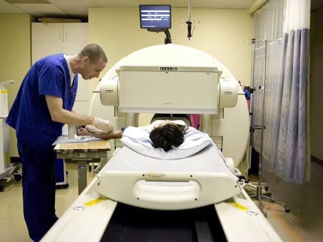
The Ventilation-Perfusion (V/Q) scan is a nuclear medicine imaging test used to evaluate the airflow (ventilation) and blood flow (perfusion) in the lungs. It plays a crucial role in the diagnosis of various pulmonary conditions, most notably pulmonary embolism (PE).
The V/Q Scan Technique – A Step-by-Step Approach
The V/Q scan consists of two main parts: the ventilation scan and the perfusion scan, which are typically performed sequentially. The fundamental principle is to introduce small amounts of radioactive tracers into the body (one inhaled for ventilation, one injected for perfusion) and use a special camera (gamma camera) to capture images that demonstrate the distribution of these tracers within the lungs.
Step 1: The Ventilation Scan (V Scan)
- Purpose: To assess how well air is moving through the airways and into the alveoli (the tiny air sacs where gas exchange occurs). Areas of the lung that are poorly ventilated will receive less of the inhaled radioactive tracer.
- Tracer: The patient inhales a small amount of a radioactive gas (commonly Xenon-133 or Krypton-81m) or a radioactive aerosol (commonly Technetium-99m DTPA or Tc-99m Technegas, which are carbon particles labelled with Tc-99m). The choice of tracer depends on availability, desired imaging technique (gas allows for washout studies), and ease of administration.
- Procedure:
- If using a gas, the patient breathes the gas mixed with air through a mouthpiece or mask while sitting or lying under the gamma camera. Images are acquired during breathing and during a “washout” phase (breathing normal air to see how quickly the gas leaves the lungs). This dynamic imaging provides insights into air trapping.
- If using an aerosol, the patient inhales the fine particles through a nebulizer and mouthpiece for several minutes. The particles deposit in the small airways and alveoli. Static images are then acquired. Aerosols are generally easier for the patient to inhale consistently and are more widely used than gases.
- Imaging: A gamma camera positioned near the chest detects the gamma rays emitted by the inhaled tracer. Multiple views (anterior, posterior, lateral, and oblique) are typically obtained to create a complete 3D representation of ventilation throughout the lungs.
Step 2: The Perfusion Scan (Q Scan)
- Purpose: To assess blood flow to the capillaries surrounding the alveoli. Areas of the lung that are poorly perfused will receive less of the injected radioactive tracer.
- Tracer: The patient receives an intravenous injection of a small amount of Technetium-99m (Tc-99m) labeled macroaggregated albumin (MAA). These are tiny particles (20-100 micrometers) that are larger than capillaries but small enough to travel through larger vessels.
- Procedure: After injection, the particles travel through the bloodstream and are distributed to the lungs proportional to regional pulmonary arterial blood flow. Because the particles are slightly larger than capillaries, they become temporarily trapped in the capillary bed. It’s important to note that only a tiny fraction (less than 0.1%) of the total pulmonary capillaries are temporarily blocked by these particles during a standard dose, making the test safe for most patients. The injection is typically given with the patient lying down to ensure even distribution of blood flow (and thus the tracer) throughout the lungs.
- Imaging: Immediately after the injection, the gamma camera is used to capture images of the chest from multiple angles (the same views as the ventilation scan) to show the distribution of the Tc-99m MAA particles, reflecting regional pulmonary perfusion.
Step 3: Image Acquisition and Processing
- Equipment: A gamma camera (planar or SPECT/CT) is used to detect the gamma radiation. Planar imaging provides 2D images from multiple angles. Single-photon emission computed tomography (SPECT) involves rotating the camera around the patient to create cross-sectional 3D images, which can improve the detection of small defects and aid in localization. SPECT/CT adds anatomical context from a simultaneous low-dose CT scan.
- Image Comparison: The core of the V/Q scan interpretation lies in comparing the ventilation images directly with the perfusion images. The images from both parts are displayed side-by-side for careful analysis, often using the same projection angles. Advanced processing software allows for quantitative assessment and enhancement of subtle findings.
Step 4: Interpretation Principles
Radiologists or nuclear medicine physicians interpret the V/Q scan images by comparing the patterns of ventilation and perfusion.
- Normal Finding: Uniform distribution of both inhaled tracer (V) and injected tracer (Q) throughout all segments and lobes of the lungs, with a V/Q ratio close to 1. This indicates normal airflow and normal blood flow that are well matched.
- Matched Defect: A decrease in both ventilation and perfusion in the same area of the lung. This pattern is typically caused by underlying lung parenchymal disease (e.g., pneumonia, COPD, asthma, pleural effusion) that affects both airflow to and blood flow within the affected region. The V/Q ratio in the affected area remains relatively normal despite the overall reduction in activity.
- Mismatch Defect: Areas of reduced or absent perfusion (Q) in segments or subsegments of the lung that are otherwise normally ventilated (V). This pattern creates a high V/Q ratio in the affected region. This is the classic finding highly suggestive of pulmonary embolism, where a clot blocks blood flow to a portion of the lung that is still receiving air normally.
- Reverse Mismatch Defect: A less common finding where there is a defect in ventilation but relatively preserved perfusion in that area. This can be seen in conditions like pulmonary edema or certain airway obstructions.
- Non-segmental Defects: Defects in perfusion that do not conform to the standard anatomical segments of the lung. These are less specific for PE and can be seen in conditions like pleural effusions or large mediastinal masses compressing vessels.
The interpretation integrates these findings with the patient’s clinical history and presentation to determine the probability of pulmonary embolism.
Role of V/Q Scan in Pulmonary Embolism (PE)
Pulmonary Embolism (PE) is a potentially life-threatening condition where a blood clot (typically from deep veins in the legs or pelvis) travels to the lungs and blocks one or more pulmonary arteries. This blockage impairs blood flow to lung tissue that is still ventilated, leading to a V/Q mismatch.
The V/Q scan is a valuable tool in the diagnostic workup for suspected PE, particularly when a V/Q mismatch pattern is identified. The classic PE finding is one or more segmental or subsegmental perfusion defects in areas with normal ventilation.
Interpreting a V/Q scan for PE often follows well-established criteria, such as those from the Prospective Investigation of Pulmonary Embolism Diagnosis (PIOPED) study, though modified criteria are now common. These criteria classify the scan results into categories reflecting the probability of PE:
- High Probability Scan: Multiple large or medium-sized segmental perfusion defects with normal ventilation in those regions (segmental V/Q mismatches). This pattern is strongly indicative of PE.
- Normal Scan: Uniform distribution of both ventilation and perfusion throughout the lungs. Rules out PE with high certainty.
- Low Probability Scan: Small, non-segmental perfusion defects, or perfusion defects matched by ventilation defects where PE is considered less likely based on the specific pattern. While PE is not excluded, the probability is low.
- Intermediate (Indeterminate) Probability Scan: Findings that do not fit into the normal, low, or high probability categories. This can include various patterns, particularly matched ventilation and perfusion defects in patients with moderate to severe lung disease, single moderate-sized mismatch defects, or difficult-to-interpret images. Indeterminate scans are common, especially in patients with underlying cardiopulmonary disease, and often require additional investigations to confirm or exclude PE.
Patients with high probability scans are usually presumed to have PE and treated accordingly. Normal scans effectively rule out PE. Patients with low or intermediate probability scans often require further testing (such as ultrasound of leg veins or CTPA, if not initially feasible) or clinical follow-up depending on their clinical pre-test probability.
Indications for V/Q Scan
While CT Pulmonary Angiography (CTPA) has become the primary imaging modality for suspected PE in many centers due to its speed and direct visualization of the thrombus, the V/Q scan remains an important and often preferred diagnostic tool in specific clinical scenarios:
- Suspected Pulmonary Embolism with Contraindications to CTPA: This is the most common indication for V/Q scanning today. Contraindications to CTPA include:
- Renal Insufficiency/Failure: CTPA requires intravenous iodinated contrast media, which is nephrotoxic. V/Q scanning uses radioactive isotopes that do not require iodinated contrast, making it safer for patients with impaired kidney function.
- Severe Allergy to Iodinated Contrast Media: Patients with a history of severe allergic reactions to contrast media are at high risk for repeated reactions with CTPA.
- Pregnancy: While radiation exposure is a concern in pregnancy for any imaging test, the total radiation dose to the fetus from a V/Q scan (particularly if a careful protocol is used, potentially omitting the ventilation phase or using specific tracers) is generally lower than that from a CTPA. The dose from the V/Q scan is primarily directed to the mother’s lungs, whereas in CTPA, scatter radiation and transit of contrast through the mother’s blood vessels contribute to fetal dose. V/Q scan is often considered the safer option in pregnant patients suspected of PE if ultrasound of leg veins is negative or inconclusive.
- Inability to Cooperate for CTPA: Patients unable to hold their breath (e.g., due to pain, anxiety, inability to follow commands) may have non-diagnostic CTPA scans. V/Q imaging is less dependent on breath-holding.
- Quantitative Assessment of Pulmonary Function: V/Q scans can be used to quantitatively assess the relative contribution of each lung or even individual lobes to total ventilation and perfusion. This is particularly useful in:
- Pre-operative Evaluation: Before lung surgery (e.g., pneumonectomy, lobectomy, lung volume reduction surgery), a quantitative V/Q scan helps predict post-operative lung function and assess surgical risk.
- Lung Transplant Evaluation: Used to assess the function of potential donor lungs or the recipient’s remaining lung tissue.
- Assessment of Chronic Thromboembolic Pulmonary Hypertension (CTEPH): V/Q scans can help identify areas of chronic pulmonary vascular obstruction in patients with suspected CTEPH, guiding further investigation and potentially surgical intervention.
- Baseline Scan: In patients with chronic lung conditions, a baseline V/Q scan might be performed for future comparison.
V/Q Scan vs. CT Pulmonary Angiography (CTPA): A Comparison
Both V/Q scan and CTPA are imaging modalities used in the evaluation of suspected PE, but they differ significantly in their technique, information provided, advantages, and disadvantages.
| Feature | V/Q Scan | CT Pulmonary Angiography (CTPA) |
|---|---|---|
| Technique | Nuclear medicine: Inhaled radioactive gas/aerosol + injected radioactive particles, detected by gamma camera. | CT scan: Intravenous iodinated contrast followed by X-ray imaging. |
| Information | Assesses functional airflow (ventilation) and blood flow (perfusion) patterns. | Assesses anatomy of the pulmonary arteries and lung parenchyma. Visualizes thrombus directly. |
| Primary Finding for PE | V/Q Mismatch (perfused defect in a ventilated area). | Direct visualization of a filling defect within a pulmonary artery. |
| Radiation Dose | Generally lower total effective dose than CTPA, especially to organs outside the chest. Fetal dose lower in pregnancy. | Usually higher total effective dose than V/Q scan. Fetal dose higher in pregnancy. |
| Contrast Medium | Radioactive isotopes (inhaled gas/aerosol, injected MAA). No iodinated contrast required. | Iodinated contrast medium required intravenously. |
| Risk of Allergic Reaction | Low risk from radiotracers. | Risk of allergic reaction, including severe anaphylaxis, to iodinated contrast. |
| Risk of Kidney Damage | Negligible risk from radiotracers. | Risk of contrast-induced nephropathy, especially in patients with pre-existing renal dysfunction. |
| Spatial Resolution | Lower resolution compared to CTPA. Difficult to detect small subsegmental clots directly. | Higher spatial resolution. Better for visualizing distal arteries and small clots. |
| Utility in Patients with Lung Disease | Interpretation is often challenging and can lead to indeterminate results due to matched defects. | Less affected by underlying lung disease, as it focuses on the vessels. Can identify other lung pathologies contributing to symptoms. |
| Speed/Availability | Can take longer to perform (30-60 mins). Availability may be limited to nuclear medicine departments. | Faster acquisition time. More widely available in emergency settings. |
| Other Findings | Primarily focused on V/Q patterns. Limited assessment of other thoracic structures. | Excellent visualization of lung parenchyma, pleura, mediastinum, chest wall. Can detect other causes of symptoms (pneumonia, effusion, etc.). |
In clinical practice, the choice between V/Q scan and CTPA for suspected PE is made based on several factors, including:
- Clinical Probability of PE: High probability warrants rapid investigation, often favoring CTPA if feasible.
- Patient Factors: Renal function, history of contrast allergy, pregnancy status, ability to cooperate.
- Availability and Expertise: Access to nuclear medicine or CT services and experienced interpreters.
- Likelihood of Underlying Lung Disease: Patients with significant COPD or other lung disease are more likely to have indeterminate V/Q scans, potentially favoring CTPA if not otherwise contraindicated.
Conclusion
The V/Q scan remains a vital diagnostic imaging modality in the evaluation of pulmonary function and, specifically, in the diagnosis of pulmonary embolism. Understanding its two-part technique – assessing ventilation by inhalation and perfusion by injection, followed by comparative imaging – is key to appreciating its utility. While CTPA offers direct visualization of clots and anatomical detail, the V/Q scan provides crucial functional information and serves as a safer alternative for patients with contraindications to iodinated contrast or specific conditions like pregnancy and renal failure. Its continued role in pre-surgical assessment and evaluation of chronic conditions further underscores its enduring value in respiratory medicine. A professional approach to interpreting V/Q scans requires careful comparison of ventilation and perfusion images in the context of the patient’s clinical presentation and relevant medical history.


