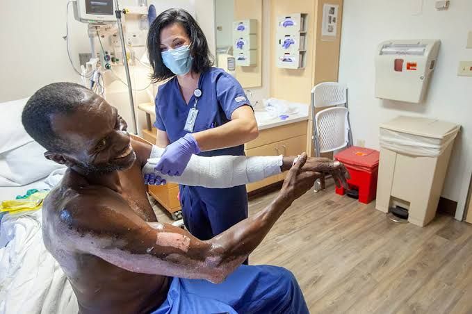
Management of Major Burns: A Comprehensive Overview
Managing a patient with major burns requires a systematic and prompt approach, starting with a thorough assessment and extending through initial resuscitation, wound care, and definitive treatment. This document outlines essential components of this process.
Relevant History for Burns
A detailed history is crucial for understanding the burn mechanism, associated injuries, and potential complications. Key elements to ascertain include:
-
- Mechanism of Injury:
- Type of Burn: Was it a flame burn (e.g., house fire, ignition of clothing), scald (e.g., hot liquid, steam), chemical burn (specify agent if known), electrical burn (high or low voltage), or radiation burn? Each type has unique characteristics and potential underlying injuries.
- Source Temperature: While often unknown precisely, understanding if the source was boiling water, hot oil, steam, or an open flame gives context to potential depth.
- Duration of Exposure: How long was the patient in contact with the heat source? Prolonged exposure, even to moderate temperatures, increases burn depth.
- Environment of Injury:
- Closed Space vs. Open Space: Burns sustained in a closed space significantly increase the risk of inhalation injury due to concentrated smoke, heat, and toxic gases. This is a critical indicator for potential airway compromise and systemic toxicity.
- Presence of Fire Retardants or Chemicals: Certain materials release toxic fumes when burned.
- Location: Was the incident at home, work, outdoors? This can provide clues about potential hazards.
- Associated Injuries:
- Trauma: Was there associated blunt trauma (e.g., fall from height while escaping fire, impact from an explosion), penetrating trauma, or fractures? Burns often occur in chaotic environments where other injuries are common.
- Blast Injury: If the burn was due to an explosion, assess for blast lung, eardrum rupture, or other blast effects.
- Inhalation Injury: Specifically inquire about symptoms like cough, hoarseness, difficulty breathing, or sputum with soot. Ask about the environment (closed space) and any loss of consciousness.
- Carbon Monoxide Exposure: Was the patient in an enclosed space with smoke? Symptoms can include headache, nausea, dizziness, confusion, and cherry-red skin (though often not present or subtle).
- Other Toxic Exposures: Were other chemicals involved in the fire?
- Patient Factors/Past Medical History:
- Pre-existing Conditions: Diabetes (affects healing, microcirculation), cardiovascular disease (affects fluid tolerance, cardiac reserve), respiratory disease (exacerbated by inhalation injury), renal disease, immunosuppression.
- Medications: Especially anticoagulants, steroids, or immunosuppressants.
- Allergies: To medications (especially pain relievers, antibiotics, anesthetics) or dressings.
- Last Tetanus Shot: Essential for wound care planning.
- Social History: Smoking history (impacts respiratory status), alcohol/drug use (affects pain management, cooperation, potential underlying issues), living situation (impacts discharge planning).
- Time Since Injury: Crucial for determining the stage of fluid resuscitation and potential for developing edema.
- Mechanism of Injury:
Burn Depth and Size in a Patient with a Major Burn
Accurate assessment of burn depth and size is fundamental for classification (minor, moderate, major), guiding initial management (fluid resuscitation), and planning definitive treatment (wound care, grafting).
-
- Burn Depth:
- Initially, depth can be challenging to assess, especially with edema or soot. Reassessment over 24-48 hours is often necessary as the burn evolves.
- Depth is described based on the layers of skin involved:
- Superficial (First-Degree): Affects only the epidermis. Appears red, dry, painful. Blanches with pressure. No blistering. Heals within 3-6 days without scarring. (Generally not included in TBSA calculations for major burns).
- Partial Thickness: Involves the epidermis and a portion of the dermis.
- Superficial Partial Thickness (Second-Degree): Affects the epidermis and papillary dermis. Appears red, moist, very painful to touch and air. Characterized by blisters. Blanches with pressure and refills quickly. Heals within 1-3 weeks with minimal scarring, though pigment changes may occur.
- Deep Partial Thickness (Deep Second-Degree): Affects the epidermis and the deeper reticular dermis. Appears pink-to-waxy white, slightly moist or dry. Pain is reduced due to nerve damage, but the patient can still feel pressure. Blisters may be present but are often ruptured or larger/flatter. Capillary refill is sluggish or absent. Healing is prolonged (3-8 weeks or longer) and typically results in hypertrophic scarring and contractures without grafting.
- Full Thickness (Third-Degree): Destroys the entire epidermis and dermis. May extend into subcutaneous tissue. Appears waxy white, leathery, dry, or charred black/brown. No pain sensation within the burn itself due to complete nerve destruction (though surrounding less deep burns are painful). Does not blanch with pressure. Requires skin grafting for healing as no epithelial cells remain. Scarring and contractures are severe.
- Fourth-Degree: Extends through skin into underlying fascia, muscle, or bone. Appears charred and involves deep structures. Requires extensive surgical reconstruction or amputation.
- Burn Size:
- Burn size is expressed as a percentage of Total Body Surface Area (% TBSA) covered by partial and full-thickness burns. Superficial burns are generally excluded from TBSA calculations for fluid resuscitation purposes, although they contribute to pain.
- For major burns, size is a primary determinant of resuscitation needs and patient outcome.
- Burn Depth:
Percentage and Degree of Burns
Accurately determining the percentage of TBSA affected by partial and full-thickness burns is critical for applying resuscitation formulas and classifying the burn severity. Assessing the degree (depth) helps predict healing time and the need for grafting.
-
- Methods for Determining TBSA:
- Rule of Nines: A quick method for adults. Divides the body into regions roughly representing 9% or multiples of 9% of the TBSA:
- Lund-Browder Chart: The most accurate method, especially for children. Uses a diagram that adjusts the relative percentage contribution of different body regions with age. The burn size is estimated on the diagram and summed. Requires tracing or drawing the burn areas.
- Palmar Method: Useful for irregular or scattered burns. The surface area of the patient’s palm plus fingers is approximately 1% of their TBSA. This can be used to estimate the size of scattered burn areas or to quickly check estimates from Rule of Nines or Lund-Browder.
- Determining Degree: This involves visual inspection and assessment of sensation, color, moisture, capillary refill, and presence/type of blisters. As noted above (Section 2), this requires experience and often reassessment.
- Methods for Determining TBSA:
Indications for Admission
Not all burn patients require hospital admission. However, patients with major burns or specific characteristics necessitating inpatient care should be admitted, often to a specialized burn center. Indications for admission include:
-
- Size of Burn:
- Partial-thickness burns > 10% TBSA (in adults).
- Any full-thickness burn (regardless of size).
- Partial-thickness burns > 5% TBSA in children or the elderly.
- Location of Burn:
- Burns involving critical areas: face, eyes, ears, hands, feet, genitalia, perineum, or major joints. These areas are at high risk for functional or cosmetic impairment.
- Mechanism of Injury:
- Inhalation injury (suspected or confirmed).
- Electrical burns (high voltage carries risk of cardiac arrhythmias, deep tissue damage, and kidney injury from myoglobinuria; low voltage may also cause significant internal injury).
- Chemical burns (especially involving strong acids or alkalis, large areas, or uncertain agents).
- Associated Injuries:
- Significant trauma accompanying the burn.
- Suspected or confirmed non-accidental trauma.
- Patient Factors:
- Pre-existing medical conditions that could complicate management or increase mortality (e.g., cardiac, pulmonary, renal disease, diabetes, immunosuppression).
- Extremes of age (infants/young children and the elderly) due to higher risk and lower physiological reserve.
- Psychiatric illness or social circumstances potentially affecting outpatient care adherence or safety.
- Pain Control:
- Burns requiring intravenous pain medication because oral analgesia is insufficient.
- Size of Burn:
Pain Management
Burn pain is often severe and multifactorial (nociceptive from tissue damage, neuropathic from nerve injury, procedural pain). Effective pain management is critical for patient comfort, cooperation with care, and reducing the stress response.
-
- Assessment: Use age-appropriate pain scales (e.g., Wong-Baker FACES, Numeric Rating Scale,oucher’s scale for non-verbal patients). Assess pain frequently, especially before and after procedures.
- Pharmacological Approaches:
- Opioids: The cornerstone for moderate to severe burn pain (e.g., IV fentanyl, morphine, hydromorphone). Titrate to effect, considering respiratory status and potential for tolerance. Patient-controlled analgesia (PCA) can be effective.
- Non-Opioids: Acetaminophen and NSAIDs (if not contraindicated by renal function or GI risk) can be used as adjuncts for mild-to-moderate pain and to reduce opioid requirements.
- Adjuvants: Benzodiazepines (e.g., midazolam, lorazepam) can help with anxiety and procedural pain. Gabapentin or pregabalin may be useful for neuropathic pain component. Ketamine can be effective for procedural pain or as an opioid-sparing agent.
- Non-Pharmacological Approaches:
- Anxiety Reduction: Creating a calm environment, providing information, presence of family.
- Distraction: Music, relaxation techniques, guided imagery, virtual reality (increasingly used in burn centers).
- Cooling: Immediate application of cool (not ice!) water can reduce pain for minor burns, but should be used cautiously in major burns to avoid hypothermia.
- Procedural Pain: Specific strategies are needed for dressing changes, physiotherapy, and other painful procedures. This often involves scheduled analgesia, anxiolysis, and potentially short-acting agents like IV fentanyl or ketamine immediately prior to the procedure.
- Addressing Underlying Issues: Treat infection, manage edema, ensure proper wound care to reduce pain.
Fluid Replacement
Fluid resuscitation is paramount in managing major burns (>15-20% TBSA in adults). The loss of capillary integrity in burned and adjacent tissues leads to massive fluid shifts from the intravascular space into the interstitial space (burn edema), causing hypovolemic shock if not corrected.
-
- Goal: Maintain adequate tissue perfusion and organ function while avoiding complications like over-resuscitation (pulmonary edema, abdominal compartment syndrome).
- Timing: Resuscitation should start as soon as possible using an intravenous line (or intraosseous if IV access is difficult).
- Fluid Type: Isotonic crystalloid solutions (e.g., Lactated Ringer’s) are the standard for initial resuscitation. Colloids (e.g., albumin) may be added later (typically after 12-24 hours) in some protocols, but their role in the initial phase is debated. Dextrose-containing fluids are generally avoided in the first 24 hours as they can contribute to hyperglycemia.
- Formulas: The Parkland formula is the most common guide:
- Total Fluid (mL) in first 24 hours = 4 mL x Patient Weight (kg) x % TBSA Burned (partial + full thickness)
- This calculated volume (usually Lactated Ringer’s) is administered over the first 24 hours from the time of burn injury.
- Administration Schedule:
- First half of the calculated volume is given over the first 8 hours from the time of injury.
- Second half of the calculated volume is given over the next 16 hours.
- Example: A 70 kg adult with 40% TBSA burn: 4 mL x 70 kg x 40 = 11,200 mL over 24 hours. 5,600 mL in the first 8 hours (700 mL/hr), 5,600 mL in the next 16 hours (350 mL/hr).
- Modification for Children: Use 3 mL/kg/% TBSA and add maintenance fluid (e.g., D5 Lactated Ringer’s at 4 mL/kg/hr for first 10 kg, + 2 mL/kg/hr for next 10 kg, + 1 mL/kg/hr for remaining weight) as children have less glycogen stores and are prone to hypoglycemia.
- Monitoring and Titration: Formulas are guides, not rigid rules. Fluid administration must be titrated based on clinical indicators of adequate resuscitation:
- Urine Output: Primary guide. Target: Adults 0.5-1 mL/kg/hr; Children < 30 kg 1 mL/kg/hr.
- Heart Rate: Should decrease towards normal range.
- Blood Pressure: Should normalize, but can be misleading due to peripheral vasoconstriction.
- Capillary Refill: Should improve.
- Mental Status: Should be alert and oriented (if not due to other injuries).
- Base Deficit/Lactate: Improving base deficit and decreasing lactate indicate improved perfusion.
- Fluid in Second 24 Hours: Typically involves colloid-containing fluids or IV fluids at potentially lower rates, guided by urine output and patient status.
Wound Management (Open, Closed, Principles of Antiseptic Solutions)
Burn wound management aims to prevent infection, promote healing (or prepare for grafting), minimize scarring, and reduce pain.
-
- Initial Management:
- Clean the wound gently with sterile saline or mild antiseptic solution.
- Debride loose skin and ruptured blisters. Intact small blisters may be left unless on areas of high friction or infection risk.
- Open Method:
- The burn wound is left exposed to air after cleaning and application of a topical antimicrobial agent.
- Advantages: Easier monitoring of the wound, potentially less painful dressing changes (no removal of adhesive or stuck dressings), allows formation of a dry eschar in deep burns.
- Disadvantages: Risk of contamination from the environment, requires a controlled environment (e.g., burn unit with laminar flow), may lead to faster evaporative heat and water loss, susceptible to trauma.
- Use: More common for facial burns (allows observation and avoids constricting dressings) and sometimes superficial burns.
- Closed Method:
- The burn wound is covered with a dressing system after cleaning and topical antimicrobial application.
- Advantages: Creates a protective barrier against infection, maintains a moist wound healing environment (beneficial for partial-thickness burns), reduces pain and evaporative loss, provides some mechanical protection.
- Disadvantages: Requires skilled application and removal, dressings need regular changes (every 1-7 days depending on material and wound status), difficult to inspect the wound without removal, potential for maceration of surrounding skin.
- Use: Most commonly used method for partial and full-thickness burns on the trunk and extremities. Various dressing types exist (e.g., gauze with topical agent, silver-impregnated dressings, hydrocolloids, synthetic materials).
- Principles of Antiseptic Solutions and Topical Antimicrobials:
- Burn wounds are highly susceptible to infection. Topical agents are essential to reduce bacterial load and prevent invasive infection. Systemic antibiotics are not routinely used prophylactically but are indicated for confirmed infection.
- Common Agents:
- Silver Sulfadiazine (Silvadene): Broad spectrum, relatively painless. Can cause transient neutropenia and is not ideal for facial burns (causes cosmetic mask) or in patients with sulfa allergy. Can penetrate eschar poorly.
- Mafenide Acetate (Sulfamylon): Good eschar penetration, useful for electrical burns or thick eschars. Can cause significant pain on application and metabolic acidosis (due to carbonic anhydrase inhibition).
- Silver Nitrate Solution: Wet dressings (e.g., 0.5%). Broad spectrum, low toxicity. Requires frequent changes (e.g., every 2 hours), stains everything black, causes electrolyte abnormalities (leaching of sodium, potassium, calcium, chloride). Poor eschar penetration.
- Povidone-Iodine: Broad spectrum. Can be painful and cause iodine toxicity (especially in large burns), impairs wound healing in high concentrations. Use is limited.
- Acticoat (Silver-impregnated dressings): Release silver ions for antimicrobial effect. Can stay in place for several days. Require wetting with sterile water.
- Bacitracin/Polymyxin B (e.g., Polysporin): Useful for superficial burns, less effective for deeper burns or established high bacterial load.
- Selection: Choice depends on burn depth, location, bacterial flora, patient allergies, and institutional protocols. Goal is to find an agent that reduces bacterial load without causing systemic toxicity or impeding wound healing.
- Initial Management:
The Value of Skin Grafting
Skin grafting is a fundamental surgical procedure in burn care, essential for the closure of deep partial-thickness and full-thickness burns that will not heal spontaneously. Its value is multifaceted:
- Allows Wound Closure: Deep burns lack sufficient remaining epithelial cells to regenerate the skin surface. Grafting provides a new epithelial layer (and dermis in full-thickness grafts or engineered skin substitutes) to close the wound.
- Prevents Infection: An open wound is a major portal for infection. Grafting closes the wound, reducing the risk of local and systemic sepsis.
- Reduces Pain: Once the wound is closed by a viable graft, the underlying nerve endings are covered, significantly reducing pain compared to an open wound.
- Minimizes Scarring and Contracture: While grafts do result in scarring, they significantly reduce the hypertrophic scarring and severe contractures that develop when large, deep burn wounds are allowed to heal secondarily or remain open. Early grafting can improve long-term function and appearance.
- Accelerates Healing Time: Grafted wounds heal much faster than deep burns attempting to heal secondarily. This reduces hospital stay and time to rehabilitation.
- Restores Function: Especially crucial for burns over joints, hands, face, and feet. Grafting allows for earlier mobilization and physiotherapy, preserving range of motion and function. Special techniques may be used for specific areas (e.g., hands require sheet grafts for better function).
- Improves Appearance: While not perfect, grafts provide a more aesthetically acceptable outcome than secondary healing of large, deep burns.
Skin grafts are typically autografts (taken from the patient’s unburned skin, usually as split-thickness meshed grafts) or, less commonly, allografts (from cadavers) or xenografts (from animals, usually pig skin) used temporarily for wound coverage. Meshing allows a small piece of donor skin to cover a larger recipient area.
In conclusion, the management of major burns is a complex, multidisciplinary process requiring prompt, skilled assessment and intervention across resuscitation, pain control, wound care, and surgical reconstruction. A systematic approach, guided by a thorough understanding of burn pathophysiology and treatment principles, is essential for optimizing patient outcomes.


