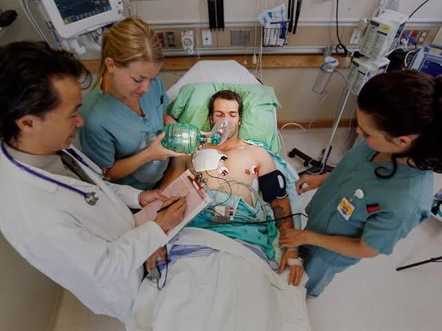
Shock represents a critical state of inadequate tissue perfusion and oxygen delivery, leading to cellular dysfunction, organ damage, and ultimately, death if not promptly corrected. It is a medical emergency requiring immediate recognition and intervention.
Defining Shock
At its core, shock is a life-threatening condition characterized by a generalized reduction in effective tissue perfusion. This deficit results in an imbalance between cellular oxygen supply and demand, leading to anaerobic metabolism, lactic acid accumulation, and progressive cellular injury. While frequently associated with low blood pressure, hypotension is a sign of shock, not its definitive definition. Tissue hypoperfusion can occur even with seemingly normal blood pressure in certain stages or types of shock. The clinical manifestations of shock arise from the body’s compensatory mechanisms and the subsequent failure of vital organs due to prolonged oxygen deprivation.
Identifying the Four Principal Categories of Shock
Shock is traditionally classified into four major categories based on the underlying pathophysiological mechanism causing inadequate tissue perfusion:
- Hypovolemic Shock: Results from a significant reduction in circulating blood volume.
- Cardiogenic Shock: Occurs when the heart fails to pump sufficient blood to meet the body’s demands.
- Distributive Shock: Characterized by widespread vasodilation, leading to a relative hypovolemia and maldistribution of blood flow. Septic shock is the most common and clinically relevant subtype of distributive shock.
- Obstructive Shock: Caused by physical obstruction of blood flow in the circulatory system or heart.
For the purpose of this guide, and aligning with common frameworks, we will focus on Hypovolemic, Cardiogenic, Septic (as the primary example of Distributive), and Neurogenic (a specific subtype of Distributive shock often considered separately due to its unique mechanisms).
Exploring the Causes of Each Shock Category
Each category of shock stems from distinct underlying issues:
- Hypovolemic Shock:
- Hemorrhage: Trauma, gastrointestinal bleeding (ulcers, varices), ruptured aneurysms, postpartum bleeding.
- Fluid Loss (Non-Hemorrhagic): Severe vomiting, diarrhea, excessive sweating, burns, third-space fluid loss (e.g., pancreatitis, bowel obstruction).
- Inadequate Fluid Intake: Severe dehydration.
- Cardiogenic Shock:
- Myocardial Infarction (Heart Attack): Damage to a significant portion of the heart muscle reduces pumping ability.
- Severe Heart Failure: End-stage cardiomyopathy, decompensated chronic heart failure.
- Arrhythmias: Extremely fast (tachycardia) or slow (bradycardia) heart rates that impair cardiac output.
- Valvular Rupture/Dysfunction: Acute mitral regurgitation, aortic stenosis.
- Septic Shock (a type of Distributive Shock):
- Bacterial Infections: Pneumonia, urinary tract infections, abdominal infections (e.g., peritonitis, appendicitis), skin/soft tissue infections.
- Fungal Infections: Especially in immunocompromised individuals.
- Viral Infections: (Less common cause compared to bacterial, but can occur).
- Typically initiated by an overwhelming systemic inflammatory response (SIRS) to an infection.
- Neurogenic Shock (a type of Distributive Shock):
- Spinal Cord Injury: Trauma (e.g., car accident, fall) above the level of T1-T4, disrupting sympathetic outflow.
- Spinal Anesthesia/Epidural Anesthesia: High levels blocking sympathetic tone.
- Severe Head Injury: Affecting the vasomotor center.
- Certain Medications or Poisons affecting the nervous system.
Contrasting the Effects on Key Organs (Heart, Kidney, Brain)
The differing etiologies of shock result in varied impacts on vital organs:
- Hypovolemic Shock:
- Heart: Initial effect is reflex tachycardia (increased heart rate) and increased contractility to maintain cardiac output. As volume depletion worsens, preload decreases significantly, eventually leading to decreased cardiac output despite the compensatory efforts.
- Kidney: Reduced renal blood flow due to decreased volume and sympathetic vasoconstriction leads to decreased glomerular filtration rate and urine output (oliguria/anuria). Risk of acute kidney injury (prerenal azotemia progressing to acute tubular necrosis).
- Brain: Reduced cerebral blood flow due to systemic hypotension and decreased cardiac output. Manifests as altered mental status, confusion, and eventually loss of consciousness.
- Cardiogenic Shock:
- Heart: Primary problem is the heart’s inability to pump effectively. Characterized by decreased contractility, often bradycardia or significant tachycardia, and increased filling pressures (high CVP/PCWP) due to forward flow failure. Myocardial ischemia may worsen the condition.
- Kidney: Similar to hypovolemic shock, reduced cardiac output leads to decreased renal perfusion, oliguria/anuria, and acute kidney injury.
- Brain: Reduced cerebral perfusion pressure due to low cardiac output and hypotension results in altered mental status, confusion, and potential coma.
- Septic Shock:
- Heart: Initially, the heart may be hyperdynamic with high cardiac output and tachycardia due to vasodilation and fluid resuscitation. However, myocardial depression often develops later, leading to reduced contractility and potentially lower cardiac output despite continued tachycardia and low systemic vascular resistance.
- Kidney: Complex effects. Can be prerenal due to relative hypovolemia, but inflammatory mediators directly damage renal tubules and interstitium (acute tubular necrosis), contributing to acute kidney injury even with adequate systemic BP.
- Brain: Septic encephalopathy is common, characterized by confusion, disorientation, and decreased level of consciousness. This is due to global hypoperfusion, inflammatory mediators affecting the blood-brain barrier, and metabolic derangements.
- Neurogenic Shock:
- Heart: Loss of sympathetic tone leads to unopposed parasympathetic activity. Characteristic findings include bradycardia (slow heart rate) and decreased contractility. Cardiac output is decreased due to low heart rate and severe vasodilation leading to low preload.
- Kidney: Reduced renal blood flow primarily due to severe systemic vasodilation and hypotension. Leads to decreased GFR, oliguria, and potential acute kidney injury.
- Brain: Cerebral blood flow is impaired due to severe hypotension. Although often the site of the cause (spinal/head injury), the effect is global hypoperfusion leading to altered mental status.
Recognizing Differentiating Hemodynamic Features, Diagnostic Tests, and Physical Findings
Distinguishing the type of shock is crucial for targeted therapy.
| Feature | Hypovolemic Shock | Cardiogenic Shock | Septic Shock | Neurogenic Shock |
|---|---|---|---|---|
| Hemodynamics | ||||
| Blood Pressure (BP) | Low | Low | Low (refractory to fluids) | Low |
| Heart Rate (HR) | High (compensatory tachycardia) | High or Low (often high, but bradycardia possible) | High (often very high) | Low (bradycardia – key finding) |
| Cardiac Output (CO) | Low | Low | High (early, hyperdynamic) or Low (late, myocardial depression) | Low |
| Systemic Vascular Resist (SVR) | High (compensatory vasoconstriction) | High (compensatory vasoconstriction) | Low (vasodilation – hallmark) | Very Low (loss of sympathetic tone) |
| Central Venous Pressure (CVP) | Low | High | Low/Normal (depends on fluid resuscitation) or High (right heart dysfunction) | Low |
| Physical Findings | ||||
| Skin | Cool, pale, clammy | Cool, pale, clammy | Warm, flushed (early, hyperdynamic) or Cool, pale (late, hypodynamic) | Warm, dry, flushed below level of injury; pale/cool above |
| Mental Status | Anxious –> Confused –> Comatose | Anxious –> Confused –> Comatose | Confused –> Disoriented –> Comatose (septic encephalopathy) | Alert (above injury) or Altered –> Comatose (below injury, or related to hypotension) |
| Urine Output | Low/Absent | Low/Absent | Low/Absent | Low/Absent |
| Other | Flat neck veins | Jugular venous distension, crackles in lungs (pulmonary edema) | Fever or hypothermia, signs of infection source (e.g., purulent sputum) | Absence of sweating below level of injury, poikilothermia (difficulty regulating temp) |
| Diagnostic Tests | ||||
| Initial | Hemoglobin/Hematocrit, Fluid balance, Physical exam | ECG, Cardiac enzymes, Chest X-ray, Echocardiogram | CBC (WBC count), Blood cultures, cultures from suspected source (urine, sputum, wound), Lactate, Inflammatory markers (CRP, Procalcitonin) | Spinal imaging (X-ray, CT, MRI), Neurological exam, Rule out other causes |
| Advanced | FAST exam (trauma), CT scan (internal bleeding) | Pulmonary Artery Catheterization (measures CVP, PCWP, CO, SVR) | Pulmonary Artery Catheterization (shows high CO, low SVR), Organ function tests (creatinine, bilirubin, liver enzymes) | Rule out other causes of distributive shock (e.g., sepsis) |
Implementing Monitoring Techniques
Effective monitoring is vital for diagnosing the type of shock, assessing response to therapy, and detecting complications.
- Basic Monitoring:
- Vital Signs: Continuous monitoring of heart rate, blood pressure (often with an arterial line for continuous, accurate measurement), respiratory rate, temperature, and oxygen saturation (pulse oximetry).
- Electrocardiogram (ECG): To detect arrhythmias, ischemia, or infarction.
- Urine Output: Measured hourly via a Foley catheter as a key indicator of renal perfusion and overall tissue perfusion. Goal often > 0.5-1 mL/kg/hr.
- Advanced Monitoring:
- Central Venous Pressure (CVP): Measured via a central venous catheter. Provides an estimate of right heart filling pressure (right ventricular preload). Useful for guiding fluid resuscitation, particularly in septic shock, but needs careful interpretation.
- Pulmonary Artery Catheter (Swan-Ganz): Allows measurement of CVP, pulmonary artery pressures, pulmonary capillary wedge pressure (PCWP – estimate of left heart preload), cardiac output (CO), and calculation of SVR. Provides detailed hemodynamic profile to differentiate shock types and guide therapy. Less commonly used now due to risks and mixed evidence on outcome benefits compared to less invasive methods.
- Arterial Lactate Levels: A marker of anaerobic metabolism and tissue hypoperfusion. Levels are often elevated in shock and trending levels is critical for assessing response to resuscitation.
- Mixed Venous (SvO2) or Central Venous (ScvO2) Oxygen Saturation: Reflects the balance between systemic oxygen delivery and consumption. Low values (e.g., <65-70%) indicate inadequate oxygen delivery relative to demand, often seen in hypovolemic and cardiogenic shock. Can be high in early septic shock due to maldistribution and impaired oxygen extraction.
- Echocardiography: Bedside ultrasound can assess cardiac function (contractility, valve function, presence of pericardial effusion) and estimate volume status. Essential in cardiogenic shock.
- Intra-abdominal Pressure Monitoring: Relevant in certain cases, like abdominal sepsis or trauma, as elevated pressure can impair venous return and organ perfusion.
Outlining General Principles of Intervention
Management of shock follows general principles of supporting circulation and oxygen delivery, while simultaneously addressing the underlying cause.
- General Principles (Applicable to all types):
- Airway, Breathing, Circulation (ABC): Ensure patent airway, adequate oxygenation, and circulatory support. Mechanical ventilation may be required.
- Fluid Resuscitation: Initial step in most shock types, aiming to restore intravascular volume and improve preload. Type and amount vary significantly by shock type.
- Pharmacologic Support: Use of vasopressors (to increase SVR and BP) and/or inotropes (to improve myocardial contractility) as needed to maintain adequate perfusion pressure and cardiac output.
- Address the Underlying Cause: This is paramount for definitive treatment.
- Monitoring and Reassessment: Continuously monitor patient response to therapy and adjust interventions accordingly.
- Type-Specific Interventions:
- Hypovolemic Shock:
- Fluid Intervention: Aggressive and rapid administration of intravenous fluids (crystalloids like Normal Saline or Lactated Ringer’s). Blood products are indicated for hemorrhagic shock (transfusions of packed red blood cells, plasma, platelets).
- Pharmacologic Intervention: Vasopressors (e.g., Norepinephrine) are used if blood pressure remains refractory despite adequate fluid resuscitation, but fluids are the primary intervention.
- Surgical/Procedural Intervention: Definitive control of bleeding (e.g., surgery to repair injured vessels, embolization, endoscopic hemostasis) or source of fluid loss.
- Cardiogenic Shock:
- Fluid Intervention: Judicious fluid administration. Excessive fluids can worsen pulmonary edema. Often requires careful titration based on filling pressures or ultrasound assessment.
- Pharmacologic Intervention: Inotropes (e.g., Dobutamine, Milrinone) to improve cardiac contractility. Vasopressors (e.g., Norepinephrine, Dopamine) to support blood pressure and ensure coronary perfusion. Diuretics may be needed to reduce pulmonary congestion if indicated.
- Surgical/Procedural Intervention: Urgent revascularization (Percutaneous Coronary Intervention – PCI, Coronary Artery Bypass Grafting – CABG) for shock due to myocardial infarction. Repair of valvular defects. Mechanical circulatory support devices (e.g., Intra-aortic Balloon Pump – IABP, Ventricular Assist Devices – VADs) can be used as bridge therapy.
- Septic Shock:
- Fluid Intervention: Initial aggressive fluid resuscitation (crystalloids) to address vasodilation and relative hypovolemia. Guided by hemodynamic targets (e.g., MAP, lactate clearance, ScvO2/CVP in some protocols).
- Pharmacologic Intervention: Early broad-spectrum antibiotics targeting likely pathogens, transitioned to specific antibiotics once cultures return. Vasopressors (Norepinephrine is first-line) to maintain Mean Arterial Pressure (MAP) typically >65 mmHg after fluid resuscitation. Inotropes (Dobutamine) may be added if cardiac output remains low despite adequate fluids and vasopressors. Steroids (hydrocortisone) considered in specific cases refractory to fluids and vasopressors.
- Surgical/Procedural Intervention: Source control is critical – drainage of abscesses, debridement of infected tissue, removal of infected devices (e.g., catheters, indwelling lines).
- Neurogenic Shock:
- Fluid Intervention: Cautious fluid administration to fill the dilated vascular space. Avoid excessive fluid as the circulatory volume itself is usually normal, and overload can occur if sympathetic tone returns. Guidance often based on CVP or response to fluid challenges.
- Pharmacologic Intervention: Vasopressors (e.g., Norepinephrine, Phenylephrine – pure alpha agonist) to increase SVR and blood pressure. Atropine or chronotropic agents (e.g., Isoproterenol, pacing) for severe bradycardia.
- Surgical/Procedural Intervention: Stabilization of spinal cord injury is paramount to prevent further neurological damage and potentially allow sympathetic tone to recover.
- Hypovolemic Shock:
Conclusion
Shock is a complex and life-threatening syndrome arising from a critical imbalance between oxygen delivery and cellular demand. Understanding the distinct pathophysiological mechanisms, clinical presentations, hemodynamic profiles, and appropriate monitoring techniques for each category (hypovolemic, cardiogenic, septic, neurogenic) is fundamental for rapid diagnosis. Timely, goal-directed intervention, including fluid resuscitation, pharmacologic support, and definitive treatment of the underlying cause, guided by continuous monitoring, is essential to restore tissue perfusion, prevent irreversible organ damage, and improve patient outcomes. The management of shock remains a cornerstone of critical care medicine.


