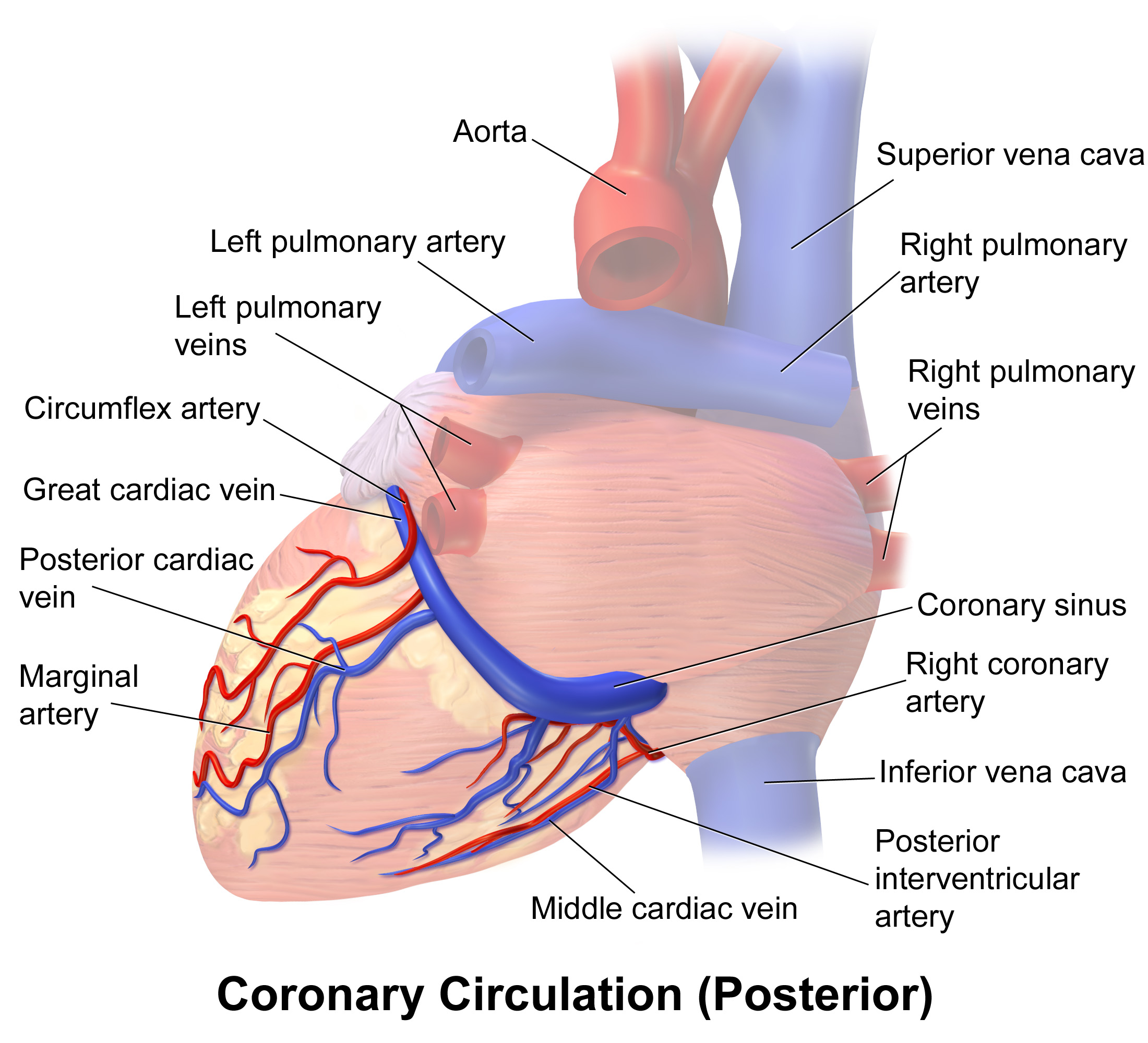
The heart, a continuously working muscle, requires a constant and substantial supply of oxygenated blood to meet its metabolic demands. This vital blood flow is provided by the coronary arteries, which branch directly from the aorta. The deoxygenated blood from the heart muscle is then collected by the cardiac veins, which primarily drain into a large vessel called the coronary sinus, ultimately returning the blood to the right atrium.
Arterial Supply: The Coronary Arteries
The coronary arteries are the first branches of the aorta, originating just above the aortic valve cusps. There are typically two main coronary arteries: the Left Coronary Artery (LCA) and the Right Coronary Artery (RCA).
1. The Left Coronary Artery (LCA)
- Origin: The LCA arises from the left aortic sinus (of Valsalva) in the wall of the ascending aorta, just superior to the left cusp of the aortic valve.
- Course: After its origin, the LCA is a short, thick vessel known as the Left Main Coronary Artery (LMCA) or Left Main Stem. It typically runs for 1-2.5 cm between the pulmonary trunk (anteriorly) and the left atrium/left auricle (posteriorly). It then usually bifurcates or occasionally trifurcates into its main branches.
- Branches and Distribution:
- Left Anterior Descending Artery (LAD): This is usually the larger of the two main terminal branches (though size varies).
- Course: The LAD travels down the anterior interventricular groove towards the apex of the heart, often curving around the apex to supply a small posterior portion.
- Branches: The LAD gives off numerous branches:
- Septal Branches: These penetrate the interventricular septum, supplying the anterior two-thirds of the interventricular septum.
- Diagonal Branches: These run diagonally across the free wall of the left ventricle, supplying the anterior and anterolateral walls of the left ventricle. Usually, there are two or three significant diagonal branches.
- Distribution: The LAD supplies the anterior wall of the left ventricle, the anterior two-thirds of the interventricular septum, the apex of the heart, and often parts of the right ventricle adjacent to the interventricular groove.
- Circumflex Artery (Cx): This is typically the other major terminal branch of the LCA.
- Course: The Circumflex artery curves posteriorly and to the left, running in the left atrioventricular groove (coronary sulcus) around the pulmonary trunk and left atrium. It extends towards the posterior side of the heart.
- Branches:
- Obtuse Marginal Branches (OM): These are usually one or more large branches that travel down the free wall of the left ventricle, supplying the lateral and posterolateral walls of the left ventricle.
- Atrial Branches: Supply the left atrium.
- Posterior Left Ventricular Branches: In cases of left dominance (discussed later), the Cx extends to the posterior interventricular groove and gives rise to the Posterior Descending Artery (PDA), supplying the inferior and posterior aspects of the left ventricle and posterior third of the septum.
- Distribution: The Cx primarily supplies the lateral and posterior walls of the left ventricle, the left atrium, and contributes to the supply of the anterolateral papillary muscle. In left-dominant systems, it also supplies the inferior wall of the left ventricle and the posterior one-third of the interventricular septum.
- Ramus Intermedius: In some cases, the LMCA trifurcates, giving rise to the LAD, Cx, and an intermediate branch (Ramus intermedius) which runs along the lateral wall of the left ventricle between the areas supplied by the LAD and Cx.
- Left Anterior Descending Artery (LAD): This is usually the larger of the two main terminal branches (though size varies).
2. The Right Coronary Artery (RCA)
- Origin: The RCA arises from the right aortic sinus (of Valsalva) in the wall of the ascending aorta, just superior to the right cusp of the aortic valve.
- Course: The RCA travels initially anteriorly, then curves to the right and posteriorly, running within the right atrioventricular groove (coronary sulcus). It continues along this groove towards the posterior aspect of the heart.
- Branches and Distribution:
- Sinus Nodal Artery: This is often the first branch, arising near the origin of the RCA (in 60% of people). It supplies the Sinoatrial (SA) node, the heart’s natural pacemaker. It courses superiorly and medial to the RCA, often looping around the superior vena cava.
- Right Marginal Artery (Acute Marginal): This is usually the largest branch arising from the free wall aspect of the RCA.
- Course: It runs along the acute margin of the right ventricle towards the apex.
- Distribution: Supplies the free wall of the right ventricle.
- Atrial Branches: Supply the right atrium.
- Atrioventricular Nodal Artery: This consistently originates from the dominant coronary artery (usually the RCA) at the ‘crux’ of the heart (where the atrioventricular and interventricular grooves meet). It supplies the Atrioventricular (AV) node.
- Posterior Descending Artery (PDA): This is a crucial branch arising at or near the crux on the posterior surface of the heart. Its origin determines coronary dominance.
- Course: It runs down the posterior interventricular groove towards the apex.
- Branches: Gives off septal branches that supply the posterior one-third of the interventricular septum, and posterolateral ventricular branches that supply the inferior and posterior walls of the left and right ventricles.
- Distribution: The PDA supplies the posterior one-third of the interventricular septum, the inferior wall of both ventricles, and the posteromedial papillary muscle of the left ventricle.
- Conus Artery: A small branch supplying the anterior wall of the right ventricle near the pulmonary conus.
Coronary Artery Dominance
The term ‘dominance’ refers to which coronary artery gives rise to the Posterior Descending Artery (PDA) and the AV nodal artery.
- Right Dominance (approx. 85%): The RCA reaches the crux and gives rise to the PDA and the AV nodal artery. This is the most common pattern.
- Left Dominance (approx. 8%): The Circumflex artery (a branch of the LCA) reaches the crux and gives rise to the PDA and the AV nodal artery.
- Co-Dominance (approx. 7%): Both the RCA and the Circumflex artery reach the crux and give off branches that run in the posterior interventricular groove, supplying the posterior septum and inferior wall. The PDA may arise from the RCA, and a large posterior left ventricular branch from the Circumflex also supplies the posterior septum and inferior wall, or a PDA may arise from both.
Sites of Anastomosis Between Branches of Coronary Arteries
Anastomoses are connections between blood vessels. While numerous small anastomoses exist between branches of the coronary arteries, they are generally considered functionally insufficient in healthy individuals to prevent myocardial infarction (heart attack) if a major coronary artery suddenly becomes blocked. However, in conditions of chronic ischemia (reduced blood flow), these anastomoses can enlarge and become more functionally significant collateral vessels.
Key areas where anastomoses occur include:
- Interventricular Septum: Septal branches of the LAD (from the LCA) anastomose with septal branches of the PDA (from the RCA or Cx, depending on dominance).
- Along the Atrioventricular Grooves: Branches of the RCA in the right AV groove anastomose with branches of the Circumflex artery in the left AV groove, particularly on the posterior surface.
- Apex: Branches of the LAD anastomose with branches of the PDA at the apex of the heart.
- Conus Artery Anastomosis: The Conus artery (from the RCA) often forms an anastomosis with the first septal perforating branch of the LAD, sometimes referred to as the “circle of Vieussens,” although its functional significance is debated.
Normal Variations in the Course of the Coronary Arteries and Their Branches
Variations in coronary anatomy are common. While many are benign, some can have clinical implications, especially during cardiac procedures or in certain pathological conditions.
Common variations include:
- Origin Variations:
- Origin of one or both coronary arteries from the pulmonary artery (Anomalous Origin of a Coronary Artery from the Pulmonary Artery – ALCAPA/ARCAPA), a serious congenital anomaly.
- Origin of the LCA and/or RCA from the opposite aortic sinus, sometimes coursing between the aorta and pulmonary trunk (interarterial course), which can lead to compression and sudden cardiac death.
- Origin of the Circumflex artery from the RCA or the right aortic sinus.
- High origin of the coronary arteries, arising above the sinotubular junction.
- Branching Pattern Variations:
- Trifurcation of the LMCA (presence of a Ramus intermedius).
- Duplication of the LAD artery.
- Variations in the origin and number of diagonal and marginal branches.
- Variations in the origin of the SA and AV nodal arteries.
- Course Variations:
- Coronary artery fistulas, abnormal connections between a coronary artery and a heart chamber or another blood vessel.
- “Myocardial bridging,” where a segment of a coronary artery (most often the LAD) tunnels through the myocardium instead of resting on the epicardial surface. This can cause compression during systole.
Venous Drainage of the Heart: The Cardiac Veins
The deoxygenated blood from the myocardium is collected by a system of cardiac veins, which largely parallel the coronary arteries. Most of these veins drain into the coronary sinus, which then empties into the right atrium. However, some veins drain directly into the heart chambers.
Major Cardiac Veins Draining into the Coronary Sinus
- Great Cardiac Vein:
- Location: It begins at the apex of the heart and ascends in the anterior interventricular groove alongside the LAD artery. It then courses posteriorly in the left atrioventricular groove alongside the Circumflex artery.
- Drainage Areas: Drains areas supplied by the LCA: the left ventricle, left atrium, and the anterior part of the interventricular septum.
- Termination: It is the main tributary of the coronary sinus and is considered the continuation of the coronary sinus at its left end.
- Middle Cardiac Vein:
- Location: It originates at the apex and ascends in the posterior interventricular groove alongside the PDA.
- Drainage Areas: Drains areas supplied by the PDA: the posterior aspects of both ventricles and the posterior part of the interventricular septum.
- Termination: Drains into the coronary sinus near its termination into the right atrium.
- Small Cardiac Vein:
- Location: It runs in the right atrioventricular groove alongside the Acute Marginal artery and the RCA.
- Drainage Areas: Drains the right ventricle and the right atrium.
- Termination: Drains into the coronary sinus, usually near its termination into the right atrium, or sometimes directly into the right atrium.
- Posterior Vein of the Left Ventricle:
- Location: Runs on the posterior surface of the left ventricle.
- Drainage Areas: Drains the posterior wall of the left ventricle.
- Termination: Drains into the coronary sinus.
- Oblique Vein of the Left Atrium (of Marshall):
- Location: A small vein descending on the posterior wall of the left atrium.
- Drainage Areas: Drains the posterior aspect of the left atrium.
- Termination: Drains into the coronary sinus. It is a remnant of the left superior vena cava.
Other Cardiac Veins Draining Directly into the Heart Chambers
- Anterior Cardiac Veins:
- Location: Several small veins running on the anterior surface of the right ventricle.
- Drainage Areas: Drain the anterior wall of the right ventricle.
- Termination: Bypass the coronary sinus and drain directly into the right atrium.
- Venae Cordis Minimae (Thebesian Veins):
- Location: Numerous tiny veins located within the walls of all four chambers.
- Drainage Areas: Drain the innermost layers of the myocardium (subendocardial region).
- Termination: Drain directly into the chambers they are located within, most numerous in the right atrium and right ventricle.
The Coronary Sinus
- Location: The coronary sinus is a large venous channel situated in the posterior part of the atrioventricular groove, between the left atrium and the left ventricle.
- Formation: It is primarily formed by the anatomical continuation of the Great Cardiac Vein as it rounds the posterior border of the heart.
- Course: It courses from left to right in the posterior atrioventricular groove, receiving tributaries along its path.
- Termination: The coronary sinus terminates by opening into the posterior wall of the right atrium, between the opening of the inferior vena cava and the tricuspid valve. The opening is typically guarded by a flap of endocardium called the valve of the coronary sinus (Valve of Thebesius).
- Tributaries: The main veins that drain into the coronary sinus are:
- Great Cardiac Vein (its origin)
- Middle Cardiac Vein
- Small Cardiac Vein
- Posterior Vein of the Left Ventricle
- Oblique Vein of the Left Atrium
Conclusion
The coronary arterial and venous systems are complex and vital for maintaining the heart’s function. The coronary arteries originate from the aorta and branch extensively to supply the myocardium. Variations in their anatomy are common. The cardiac veins primarily drain into the coronary sinus, which empties into the right atrium, completing the cycle of blood flow to the heart muscle. A comprehensive understanding of this anatomy is essential for the diagnosis and management of cardiovascular diseases.


