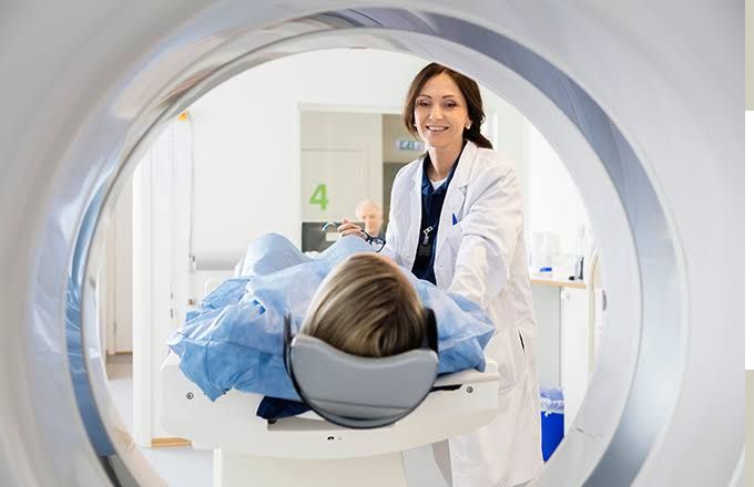
Nuclear medicine, a specialized field of medical imaging, offers unique insights into various bodily functions. Among its many applications, bone scans are instrumental in diagnosing conditions related to bone health, particularly conditions associated with osteoblastic lesions, metastatic diseases, and other skeletal abnormalities. This guide aims to enhance your understanding of nuclear medicine’s role in bone health through a structured, step-by-step approach.
Understanding Osteoblastic Lesions and Bone Reactions
What are Osteoblastic Lesions?
Osteoblastic lesions are areas of increased bone formation often seen in response to various insults to the bone, including infection, inflammation, or metastasis. In simpler terms, when the bone experiences damage or disease, specialized cells called osteoblasts respond by producing new bone material. This reaction can be visualized and analyzed through imaging techniques like bone scans.
The Bone’s Response to Insult
When the bone suffers an injury or undergoes a pathological process, the body initiates a complex healing mechanism. The stages can be summarized as follows:
- Initial Injury: Various factors can lead to a bone insult, including trauma, infections (such as osteomyelitis), and malignancies that metastasize to bone.
- Inflammatory Response: Following injury, the body’s immune system triggers an inflammatory response. The influx of inflammatory cells signals osteoblasts to increase bone production.
- Bone Formation: Osteoblasts synthesize new bone matrix, leading to an area of increased radioactivity on a bone scan, indicative of osteoblastic activity.
- Bone Remodeling: The new bone undergoes remodeling, where it may revert to a quiescent state or remain active depending on the ongoing pathological process.
Understanding this dynamic is vital as it helps medical professionals discern the underlying causes of osteoblastic lesions and their implications.
The Technique of a Bone Scan
What is a Bone Scan?
A bone scan is a nuclear imaging technique that employs small amounts of radioactive material (radiotracers) to visualize bone metabolism. The procedure is particularly useful in identifying bone diseases and conditions that disrupt normal bone function.
The Bone Scan Procedure
- Preparation: Patients are typically advised to hydrate well before the procedure and refrain from strenuous exercise or recent bone surgeries that may affect results.
- Administration of Radiotracer: A radiotracer, commonly technetium-99m (Tc-99m), is injected intravenously. The tracer has a high affinity for areas of increased bone metabolism.
- Waiting Period: After injection, patients usually rest for 2-4 hours to allow sufficient time for the tracer to accumulate in the bones.
- Imaging: The actual imaging is performed using a gamma camera, which detects gamma emissions from the radiotracer. The camera produces images that highlight areas of abnormal bone activity, appearing as “hot spots” where elevated metabolic activity is present.
- Interpretation: A nuclear medicine physician interprets the scan. Areas with increased uptake may indicate osteoblastic lesions, metastatic disease, or other abnormalities, while reduced uptake can indicate osteolytic lesions or degenerative changes.
Benefits of Bone Scans
Bone scans are non-invasive and provide a comprehensive view of skeletal health, making them invaluable in detecting:
The Role of Bone Scans in Metastasis
Bone metastasis occurs when cancer cells spread from their original site to the bone. This is particularly common with cancers such as breast, prostate, and lung cancer. Bone scans play a critical role in identifying such metastases:
- Detection of Bone Metastases: The high sensitivity of bone scans allows for the early detection of metastatic lesions, which is crucial for timely intervention.
- Evaluating Treatment Efficacy: Serial bone scans can be useful in monitoring the response to treatment plans involving chemotherapy or radiation therapy and determining whether the metastatic lesions are responding to therapy.
- Prognostic Indicator: The extent and severity of bone lesions often correlate with the prognosis of metastatic disease, providing essential information for devising patient management strategies.
Differential Diagnosis of Active Lesions on Bone Scans
Active lesions on bone scans can arise from various conditions, making differential diagnosis essential for effective patient management. Some of the most common differential diagnoses include:
- Osteoblastic Metastases: As mentioned, metastatic cancer can lead to osteoblastic lesions, particularly evident in prostate and breast cancers.
- Paget’s Disease of Bone: This chronic disorder results in abnormal bone remodeling and can be seen as areas of increased uptake on bone scans.
- Bone Infections (Osteomyelitis): This condition can also cause increased radiotracer uptake due to a vigorous inflammatory response.
- Fractures: Traumatic or stress fractures often lead to increased metabolic activity in the affected area, appearing as hot spots.
- Bone Tumors: Primary bone tumors, whether benign or malignant, can exhibit similar manifestations on scintigraphy.
- Avascular Necrosis: This condition, resulting from compromised blood supply to the bone, can also show increased uptake in the associated areas.
- Hyperparathyroidism: Increased osteoblastic activity can arise due to hyperparathyroidism, manifesting as areas of increased radiotracer uptake.
Conclusion
Nuclear medicine, through the application of bone scans, provides detailed insight into the bone’s reaction to various insults, enabling timely and precise diagnostics. From understanding processes leading to osteoblastic lesions to recognizing the utility of bone scans in assessing metastases and differentiating lesions, the significance of this imaging technique cannot be overstated. With ongoing advances in nuclear medicine, future developments will likely enhance our ability to diagnose and manage bone health, ultimately improving patient outcomes. Remember, a thorough understanding of the principles and interpretations associated with nuclear medicine is vital for healthcare professionals in delivering effective patient care.


