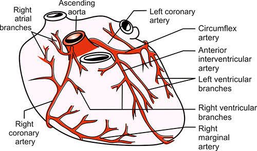
The heart is a remarkable organ requiring a constant and robust supply of oxygenated blood to perform its vital work of pumping blood throughout the body. This critical supply is delivered via the coronary arteries. Understanding the dynamics of coronary blood flow, its regulation, and the consequences of impaired flow is fundamental to comprehending cardiovascular health and disease.
Normal Coronary Blood Flow During the Cardiac Cycle
The coronary arteries originate from the aorta, just beyond the aortic valve. Unlike the systemic circulation, blood flow through the coronary arteries is highly influenced by the mechanical activity of the heart muscle itself.
- Flow During Systole (Heart Contraction): During ventricular systole, the heart muscle (myocardium) contracts forcefully. This contraction compresses the small blood vessels (capillaries and small arteries) that run within the myocardial wall. This compression significantly impedes blood flow, particularly in the deeper layers of the ventricular wall (the subendocardium). Consequently, coronary blood flow to the left ventricle is substantially reduced during systole, especially compared to diastole. The right ventricle’s wall is thinner, so systolic compression is less pronounced, resulting in a less drastic reduction in right coronary flow during systole.
- Flow During Diastole (Heart Relaxation): During ventricular diastole, the heart muscle relaxes. The compression on the coronary vessels is released, and the aortic pressure (specifically, the diastolic pressure) drives blood into the coronary arteries and through the myocardial capillary network. Therefore, the majority of blood flow to the left ventricle occurs during diastole. This reliance on diastolic pressure highlights why maintaining adequate diastolic blood pressure is crucial for proper coronary perfusion.
- Flow Distribution: Blood flow is not uniform across all layers of the myocardial wall. The subendocardial layer (the inner layer closest to the ventricular cavity) is particularly vulnerable. During systole, it experiences the greatest compressive forces, leading to the most significant reduction in flow. While flow recovers during diastole, under conditions of increased heart rate (shortening diastole) or reduced perfusion pressure, the subendocardium is the first region to suffer from inadequate blood supply. The epicardial layer (the outer layer) experiences less compression and maintains a relatively steadier flow profile throughout the cardiac cycle.
In summary, normal left ventricular coronary blood flow is predominantly diastolic, a unique characteristic necessitated by myocardial contraction, while right ventricular flow is more uniform.
Local Control of Coronary Blood Flow: Metabolism as the Primary Factor
The coronary circulation possesses remarkable autoregulatory capabilities, ensuring that blood supply closely matches the heart muscle’s metabolic demands. Local factors are the primary regulators of coronary blood flow, with metabolic activity being the most significant.
- Metabolic Regulation: The heart is almost entirely aerobic, meaning it relies heavily on oxygen to produce ATP, the energy currency for contraction. Therefore, blood flow must increase proportionally to the myocardium’s oxygen consumption.
- When myocardial activity increases (e.g., during exercise, stress), the heart muscle consumes more oxygen and produces more metabolic byproducts.
- These metabolic byproducts, such as adenosine, lactic acid, carbon dioxide, hydrogen ions, potassium ions, and others, act as potent vasodilators when released into the interstitial fluid surrounding the coronary resistance vessels (arterioles).
- The increased concentration of these vasodilators causes the arterioles to relax and dilate, increasing coronary blood flow.
- Conversely, when myocardial activity decreases, less oxygen is consumed, fewer vasodilatory metabolites are produced, leading to vasoconstriction and reduced blood flow.
- Oxygen Demand as the Primary Driver: Because the heart extracts a very high percentage (around 70-80%) of the oxygen available in the blood passing through the coronary arteries even at rest, it cannot significantly increase oxygen supply by increasing extraction, unlike many other tissues. The only practical way to increase oxygen supply to the myocardium is by increasing blood flow. Thus, myocardial oxygen demand is the primary physiological driver of coronary blood flow. Any increase in heart rate, contractility, or wall tension (factors increasing oxygen demand) must be met by a corresponding increase in coronary flow, mediated primarily by the metabolic feedback loop.
- Other Local Factors: The endothelium lining the coronary vessels also plays a role by releasing vasodilating substances like nitric oxide (NO) and prostacyclin, and vasoconstricting substances like endothelin. While important, these endothelial factors typically modulate the flow responses primarily driven by metabolic needs, especially under physiological conditions.
The Effect of the Autonomic Nervous System on Coronary Arteries
The autonomic nervous system (ANS) also influences coronary blood flow, but its role is more complex and often indirect compared to local metabolic control. Both sympathetic and parasympathetic divisions innervate the coronary vessels.
- Sympathetic Nervous System:
- Coronary arteries contain both Alpha-1 and Beta-2 adrenergic receptors.
- Alpha-1 Receptors: Stimulation of Alpha-1 receptors leads to vasoconstriction. These receptors are more prevalent in the larger epicardial coronary arteries.
- Beta-2 Receptors: Stimulation of Beta-2 receptors leads to vasodilation. These receptors are more prevalent in the smaller resistance vessels (arterioles) within the myocardial wall.
- Sympathetic activation (e.g., during exercise or stress) directly stimulates both receptor types simultaneously.
- However, sympathetic stimulation also increases heart rate and contractility, which dramatically increases myocardial metabolic activity and oxygen demand.
- The potent local metabolic vasodilation triggered by increased oxygen demand typically overrides the direct vasoconstrictive effect of Alpha-1 receptors.
- Furthermore, Beta-2 receptor stimulation contributes to vasodilation in the resistance vessels, aiding the metabolic response.
- Therefore, the net effect of sympathetic activation under normal physiological conditions is usually a significant increase in coronary blood flow, driven primarily by the metabolic needs induced by increased cardiac work, augmented by Beta-2 mediated vasodilation, and overcoming Alpha-1 mediated constriction. Direct Alpha-1 vasoconstriction might be more significant during periods of low metabolic demand or in pathologically altered vessels.
- Parasympathetic Nervous System:
- Parasympathetic (vagal) innervation to the coronary arteries is less dense than sympathetic innervation.
- Acetylcholine, the neurotransmitter, binds to muscarinic receptors (primarily M2 receptors) on the coronary vessels.
- Stimulation can lead to vasodilation, often mediated by the release of nitric oxide from the endothelium.
- However, the primary effect of parasympathetic stimulation is to decrease heart rate and contractility, thereby reducing myocardial metabolic demand.
- Consequently, the main influence of parasympathetic activity on coronary flow is typically indirect, resulting in decreased flow due to reduced metabolic needs, rather than a strong direct vasodilatory effect.
In essence, while the ANS can directly influence coronary vessel tone, its most significant impact on flow is often indirect, by altering cardiac work and thus myocardial metabolic demand, which is the dominant regulator.
Ischemic Heart Disease, Cardiac Pain, and Collateral Circulation
When the demand for oxygen by the myocardium exceeds the supply delivered by the coronary arteries, the heart muscle becomes ischemic (oxygen-deprived). This state is known as Ischemic Heart Disease (IHD) or Coronary Artery Disease (CAD) when caused by blockages in the arteries.
- Cause of Ischemic Heart Disease: The most common cause of IHD is atherosclerosis, the buildup of plaque within the walls of the coronary arteries. These plaques narrow the arterial lumen, reducing blood flow. This reduction in flow becomes particularly problematic during periods of increased oxygen demand (like exercise), leading to a supply-demand mismatch. If a plaque ruptures, it can trigger the formation of a blood clot (thrombus), which can acutely and severely obstruct blood flow.
- Cause of Cardiac Pain (Angina Pectoris): Cardiac pain, commonly known as angina pectoris, is a hallmark symptom of myocardial ischemia. The pain is not typically caused by the lack of oxygen itself, but rather by the accumulation of metabolic byproducts in the ischemic tissue. When oxygen is insufficient, the heart muscle switches to anaerobic metabolism, which produces lactic acid. Along with other substances released by ischemic cells (such as adenosine, bradykinin, serotonin), lactic acid irritates nerve endings in the myocardium. These pain signals are transmitted via sympathetic afferent fibers to the spinal cord (segments T1-T4/T5) and then to the brain, where they are perceived as pain. The pain is often described as pressure, tightness, squeezing, or heaviness in the chest, and can radiate to the left arm, neck, jaw, or back.
- Mechanism of Collateral Circulation: The coronary artery system is not simply a set of isolated vessels. There are pre-existing, small interconnections (anastomoses) between branches of the same coronary artery and between different coronary arteries. If a major coronary artery gradually narrows over time (due to progressive atherosclerosis), the reduced blood flow in the region supplied by that artery creates a persistent state of mild chronic ischemia. This chronic ischemia acts as a stimulus that promotes the growth and enlargement of these pre-existing collateral vessels. This development of collateral circulation provides alternative routes for blood to reach the ischemic myocardial tissue, bypassing the blocked or narrowed main artery. Collateral vessels can develop over weeks or months and can provide a degree of protection, potentially reducing the severity of ischemia during increased demand or limiting the extent of damage during an acute blockage (like a heart attack), though they are often insufficient to fully restore normal flow, especially under stress.
Diagnosis of Coronary Artery Disease, Angina Pectoris, and Myocardial Infarction
Diagnosing coronary artery disease and its manifestations (angina and myocardial infarction) involves integrating patient history, physical examination, and a range of diagnostic tests.
- Coronary Artery Disease (CAD): CAD is the underlying structural disease. Diagnosing CAD involves assessing the presence and extent of atherosclerotic plaques in the coronary arteries.
- Assessment of Risk Factors and Symptoms: Evaluating patient history for cardiovascular risk factors (hypertension, high cholesterol, diabetes, smoking, family history) and symptoms suggestive of ischemia (chest pain, shortness of breath, fatigue).
- Non-Invasive Tests:
- Electrocardiogram (ECG): Can show signs of past or present ischemia or infarction, though a normal resting ECG does not rule out CAD.
- Stress Testing: Evaluates the heart’s function under increased demand (usually exercise or pharmacologically induced stress). Stress ECG, Stress Echocardiography (looks for wall motion abnormalities), and Myocardial Perfusion Imaging (MPI, nuclear stress test, looks for areas of reduced blood flow) are common types. These tests assess the functional significance of blockages.
- Coronary Calcium Scoring (CT Scan): Measures the amount of calcified plaque in the coronary arteries, indicating the presence and burden of atherosclerosis, but not the degree of narrowing.
- Coronary CT Angiography (CTA): Uses contrast dye and CT imaging to visualize the coronary arteries directly, assessing the presence and severity of stenoses (narrowings).
- Invasive Test:
- Coronary Angiography (Cardiac Catheterization): Considered the gold standard for defining coronary anatomy. A catheter is inserted through an artery (usually in the wrist or groin) and guided to the coronary arteries. Contrast dye is injected, and X-ray images (angiograms) are taken to visualize blockages. This test is also therapeutic, allowing for procedures like angioplasty and stenting.
- Angina Pectoris: Angina is a clinical syndrome characterized by chest discomfort caused by transient myocardial ischemia. Diagnosis is primarily based on:
- Patient History: A detailed description of symptoms (character, location, duration, precipitating factors like exertion, relieving factors like rest or nitroglycerin). Typical angina is predictably brought on by exertion and relieved by rest. Atypical angina may have different triggers or characteristics. Unstable angina is new-onset angina, angina that occurs at rest, or a worsening pattern of previously stable angina, indicating a higher risk of MI.
- ECG during an episode: May show transient ST-segment depression or T-wave inversion during an anginal episode, indicating ischemia. A normal ECG does not rule out angina.
- Stress Testing: As described above, useful for reproducing symptoms and showing objective signs of ischemia during stress.
- Myocardial Infarction (MI – “Heart Attack”): MI occurs when there is prolonged and severe myocardial ischemia, leading to irreversible damage and necrosis (death) of heart muscle tissue. Diagnosis is based on a combination of:
- Clinical Presentation: Severe, prolonged chest pain (often similar to angina but more intense and not relieved by rest or nitroglycerin), shortness of breath, sweating, nausea, fatigue.
- Electrocardiogram (ECG): Shows characteristic changes depending on the type and location of the infarct. ST-segment elevation (STEMI) indicates a complete blockage and requires urgent reperfusion. Non-ST-segment elevation (NSTEMI) indicates partial or fluctuating blockage. Q waves can develop hours to days later, indicating past infarction.
- Cardiac Biomarkers: Proteins released into the bloodstream from damaged heart muscle cells. Cardiac Troponins (Troponin I and Troponin T) are the most specific and sensitive markers and are the current standard for diagnosing MI. Levels rise within a few hours of injury and remain elevated for several days. CK-MB was previously used but is less specific than troponins.
In summary, CAD is the underlying condition, Angina is a symptom of reversible ischemia, and MI is the result of irreversible damage from prolonged ischemia. Diagnosis involves assessing the presence and severity of blockages (CAD diagnosis) and evaluating the clinical presentation, ECG changes, and cardiac biomarkers to determine if a patient is experiencing stable angina, unstable angina, or a myocardial infarction.


