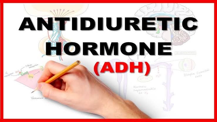
Physiological Effects of Antidiuretic Hormone (ADH)
Antidiuretic hormone (ADH), also known as vasopressin, plays a critical role in maintaining water balance and regulating blood pressure. Its primary physiological effects include:
- Water Reabsorption in the Kidneys: ADH acts on the kidneys, specifically targeting the collecting ducts and distal tubules. It increases the permeability of these structures to water by promoting the insertion of aquaporin-2 channels into their membranes. This allows more water to be reabsorbed from the filtrate back into the bloodstream, reducing urine output and conserving body water.
- Regulation of Blood Osmolarity: By increasing water reabsorption, ADH helps lower blood osmolarity when it is elevated due to dehydration or high salt intake. This ensures that solute concentrations in the blood remain within a normal range.
- Vasoconstriction: At higher concentrations, ADH causes constriction of blood vessels (vasoconstriction), which increases peripheral resistance and raises blood pressure. This effect is particularly important during significant fluid loss or hemorrhage.
- Role in Homeostasis: ADH contributes to overall homeostasis by balancing fluid levels, preventing dehydration, and maintaining stable blood pressure under varying physiological conditions.
Regulation of Antidiuretic Hormone Secretion
The secretion of ADH is tightly regulated by several factors to maintain fluid balance and osmolarity:
- Osmoreceptors in the Hypothalamus:
- Specialized osmoreceptors located in the hypothalamus monitor changes in plasma osmolarity.
- When plasma osmolarity increases (e.g., during dehydration or after consuming salty foods), these osmoreceptors shrink due to water loss, triggering signals for increased ADH release from the posterior pituitary.
- Conversely, when plasma osmolarity decreases (e.g., after drinking large amounts of water), osmoreceptors swell, leading to reduced ADH secretion.
- Baroreceptors and Blood Volume/Pressure Changes:
- Baroreceptors located in the carotid sinus and aortic arch detect changes in blood volume or pressure.
- A drop in blood volume or pressure stimulates increased ADH release to promote water retention and restore circulatory volume.
- Conversely, an increase in blood volume suppresses ADH secretion.
- Other Stimuli Influencing ADH Secretion:
- Stress, pain, nausea, hypoglycemia, and certain medications can stimulate ADH release.
- Alcohol inhibits ADH secretion, leading to increased urine production and potential dehydration.
- Feedback Mechanism: The regulation of ADH operates via a negative feedback loop:
- Increased water reabsorption lowers plasma osmolarity and restores normal hydration levels.
- Once homeostasis is achieved, osmoreceptors signal for reduced ADH secretion.
Major Physiological Effects of Oxytocin
Oxytocin has several key physiological effects primarily related to reproduction and social bonding:
- Uterine Contractions During Labor: Oxytocin stimulates powerful contractions of uterine smooth muscle during childbirth. This process is part of a positive feedback loop known as the Ferguson reflex—cervical stretching triggers oxytocin release, which intensifies contractions until delivery occurs.
- Milk Ejection Reflex (Let-Down Reflex): During breastfeeding, oxytocin causes contraction of myoepithelial cells surrounding milk ducts in the mammary glands. This leads to milk ejection into the infant’s mouth upon suckling stimulation.
- Maternal Bonding and Social Behavior: Oxytocin plays a role in maternal behaviors by fostering bonding between mother and child after birth. Additionally, oxytocin influences social bonding more broadly by promoting feelings of trust, empathy, and attachment between individuals.
- Role in Male Reproductive Function: In males, oxytocin facilitates sperm transport during ejaculation by contracting smooth muscle within reproductive ducts such as the vas deferens.
Regulation of Oxytocin Secretion
The regulation of oxytocin secretion involves specific stimuli that trigger its release through positive feedback mechanisms:
- Cervical Stretching During Labor (Ferguson Reflex):
- As labor progresses and cervical dilation occurs due to fetal movement toward the birth canal, sensory neurons send signals to the hypothalamus.
- The hypothalamus responds by stimulating oxytocin release from the posterior pituitary.
- Increased oxytocin further enhances uterine contractions until childbirth is complete.
- Suckling Stimulus During Breastfeeding:
- Stimulation of sensory receptors in the nipples during breastfeeding sends afferent signals to the hypothalamus.
- In response, oxytocin is released into circulation from axon terminals within the posterior pituitary.
- This triggers contraction of myoepithelial cells around mammary alveoli for milk ejection.
- Positive Feedback Mechanisms: Both uterine contractions during labor and milk ejection rely on positive feedback loops:
- For example:
- Uterine contractions lead to cervical stretching → more oxytocin release → stronger contractions → further cervical dilation until delivery occurs.
- Infant suckling stimulates nipple receptors → more oxytocin release → enhanced milk ejection until feeding ends.
- For example:
- Termination Signals for Oxytocin Release: Once childbirth concludes or breastfeeding ceases temporarily (e.g., between feedings), sensory input diminishes significantly—halting further oxytocin secretion until new stimuli arise again.


