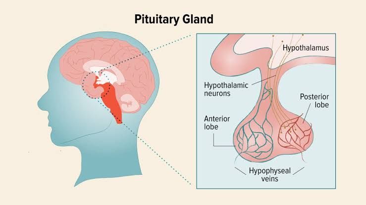
Neoplasms of the Anterior Pituitary and Their Clinical Syndromes
The anterior pituitary gland is a critical endocrine organ responsible for producing hormones that regulate various physiological processes. Neoplasms of the anterior pituitary, commonly referred to as pituitary adenomas, are benign tumors that arise from the hormone-secreting cells of the anterior pituitary. These adenomas can be classified based on their size (microadenomas <10 mm, macroadenomas ≥10 mm) and their functional status (hormone-secreting or non-functioning).
Functional Adenomas
- Prolactinomas: These are the most common functional pituitary adenomas. They secrete prolactin, leading to hyperprolactinemia. Clinical features include galactorrhea, amenorrhea in women, hypogonadism in men, infertility, and decreased libido.
- Growth Hormone-Secreting Adenomas: These cause acromegaly in adults and gigantism in children due to excessive growth hormone (GH) secretion. Symptoms include enlarged hands and feet, facial changes (e.g., prognathism), joint pain, and metabolic disturbances.
- Adrenocorticotropic Hormone (ACTH)-Secreting Adenomas: These lead to Cushing’s disease by causing overproduction of cortisol from the adrenal glands. Symptoms include central obesity, moon facies, purple striae, hypertension, and glucose intolerance.
- Thyroid-Stimulating Hormone (TSH)-Secreting Adenomas: Rarely occurring tumors that cause secondary hyperthyroidism with symptoms such as weight loss, heat intolerance, tachycardia, and tremors.
- Gonadotropin-Secreting Adenomas: These are rare functional adenomas that secrete luteinizing hormone (LH) or follicle-stimulating hormone (FSH). They may present with menstrual irregularities or hypogonadism.
Non-Functioning Adenomas
Non-functioning adenomas do not secrete active hormones but can cause symptoms due to mass effect:
- Headaches
- Visual field defects (classically bitemporal hemianopia due to compression of the optic chiasm)
- Hypopituitarism due to compression of normal pituitary tissue.
Causes and Clinical Entities Related to Hypopituitarism
Hypopituitarism refers to partial or complete deficiency of one or more anterior or posterior pituitary hormones.
Causes
- Primary Causes:
- Pituitary adenoma (most common)
- Pituitary apoplexy (hemorrhage into a tumor)
- Inflammatory conditions like lymphocytic hypophysitis
- Radiation therapy targeting the sellar region
- Genetic mutations affecting pituitary development.
- Secondary Causes:
Clinical Entities
The clinical manifestations depend on which hormones are deficient:
- Growth Hormone Deficiency: Short stature in children; reduced muscle mass and increased fat deposition in adults.
- ACTH Deficiency: Fatigue, hypotension, weight loss.
- TSH Deficiency: Features of hypothyroidism such as cold intolerance and bradycardia.
- Gonadotropin Deficiency: Amenorrhea/infertility in women; decreased libido/erectile dysfunction in men.
- Antidiuretic Hormone Deficiency: Leads to diabetes insipidus.
Diabetes Insipidus and Syndrome of Inappropriate Antidiuretic Hormone Secretion
Diabetes Insipidus (DI)
DI is characterized by an inability to concentrate urine due to insufficient antidiuretic hormone (ADH) secretion or renal insensitivity to ADH.
- Types:
- Central DI: Caused by damage to the hypothalamus or posterior pituitary leading to ADH deficiency.
- Nephrogenic DI: Due to renal resistance to ADH action.
- Clinical Features:
- Polyuria (>3 liters/day)
- Polydipsia
- Hypernatremia if water intake is inadequate.
- Diagnosis:
- Low urine osmolality (<300 mOsm/kg) despite hypernatremia.
- Water deprivation test showing failure to concentrate urine.
- Treatment:
- Central DI: Desmopressin replacement therapy.
- Nephrogenic DI: Thiazide diuretics and dietary sodium restriction.
Syndrome of Inappropriate Antidiuretic Hormone Secretion (SIADH)
SIADH involves excessive secretion of ADH leading to water retention without appropriate stimuli.
- Causes:
- CNS disorders like stroke or trauma.
- Malignancies such as small-cell lung carcinoma.
- Medications like selective serotonin reuptake inhibitors (SSRIs).
- Clinical Features:
- Hyponatremia with low plasma osmolality (<275 mOsm/kg).
- Euvolemic status without edema.
- Diagnosis:
- Urine osmolality >100 mOsm/kg despite hyponatremia.
- Treatment:
- Fluid restriction.
- Demeclocycline or vasopressin receptor antagonists for refractory cases.
Definition of Craniopharyngioma
Craniopharyngioma is a rare congenital epithelial tumor arising along the craniopharyngeal duct pathway near the sellar region.
Characteristics
- It accounts for 2–4% of all intracranial tumors.
- Commonly presents with visual disturbances due to optic chiasm compression, endocrine dysfunction from hypothalamic-pituitary axis involvement, and hydrocephalus caused by obstruction.
Pathogenesis
Craniopharyngioma originates from remnants of Rathke’s pouch during embryonic development.
Treatment
Surgical resection remains the primary treatment modality often followed by radiotherapy for residual disease control. However, long-term complications include persistent endocrine deficits requiring lifelong hormonal replacement therapy.


