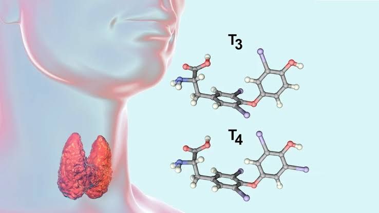
Thyroid Hormone Biosynthesis: Monoiodotyrosines, Diiodotyrosines, T3, T4, and Reverse T3
Thyroid hormone biosynthesis occurs in the thyroid gland’s follicular cells and involves several critical steps. The process begins with the production of thyroglobulin, a glycoprotein rich in tyrosine residues, which serves as the backbone for thyroid hormone synthesis. Iodide from the bloodstream is actively transported into the follicular cells and oxidized to iodine by thyroid peroxidase (TPO). This iodine is then incorporated into tyrosine residues on thyroglobulin to form monoiodotyrosine (MIT) and diiodotyrosine (DIT).
- Monoiodotyrosines (MIT) and Diiodotyrosines (DIT):
- MIT is formed when one iodine atom binds to a tyrosine residue.
- DIT is formed when two iodine atoms bind to a tyrosine residue.
- Formation of T3 and T4:
- Two DIT molecules combine to form tetraiodothyronine (T4 or thyroxine).
- One MIT molecule combines with one DIT molecule to form triiodothyronine (T3).
- Reverse T3 (rT3):
- Reverse T3 is an inactive metabolite produced by peripheral deiodination of T4. It differs structurally from active T3 because it has two iodine atoms on the outer ring and only one on the inner ring.
These hormones are stored within the colloid as part of thyroglobulin until they are cleaved and released into circulation upon stimulation by thyroid-stimulating hormone (TSH).
Metabolism of Iodide and Iodine
Iodide metabolism begins with dietary intake, where iodide is absorbed in the gastrointestinal tract and enters the bloodstream. The thyroid gland actively concentrates iodide through a sodium/iodide symporter located on the basolateral membrane of follicular cells. This process, known as “iodide trapping,” ensures that iodide levels within the thyroid gland are significantly higher than in plasma.
Once inside the follicular cell:
- Iodide is transported into the colloid via pendrin.
- Thyroid peroxidase oxidizes iodide into iodine, which can then be used for organification—binding iodine to tyrosyl residues on thyroglobulin.
Excess iodinated compounds or unused iodide can be dehalogenated by dehalogenase enzymes within follicular cells, allowing recycling of iodide for further hormone synthesis.
Role of Peroxidase, Iodinase, Coupling, Protease, Dehalogenase, and Thyroglobulin
- Thyroid Peroxidase (TPO):
- Catalyzes three key reactions:
- Oxidation of iodide to iodine.
- Organification: Binding iodine to tyrosyl residues on thyroglobulin to form MITs and DITs.
- Coupling: Combining MITs and DITs to produce T3 and T4.
- Catalyzes three key reactions:
- Iodinase:
- Refers broadly to enzymes involved in adding or removing iodine during hormone synthesis or metabolism.
- Coupling:
- A critical step where MITs and DITs are enzymatically joined by TPO to form either T3 or T4.
- Proteases:
- Enzymes within lysosomes that cleave thyroglobulin during endocytosis to release free T3 and T4 into circulation.
- Dehalogenase:
- Recycles unused or excess MITs/DITs by removing their iodine atoms so that they can be reused for new hormone synthesis.
- Thyroglobulin:
- A glycoprotein synthesized by follicular cells; it serves as both a storage matrix for thyroid hormones within colloid and a precursor for their synthesis.
Thyroid-Stimulating Hormone Action via cAMP
TSH binds to its receptor on thyroid follicular cells’ membranes, activating adenylate cyclase through G-protein signaling pathways:
- Adenylate cyclase converts ATP into cyclic AMP (cAMP), which acts as a second messenger.
- cAMP activates protein kinase A (PKA), leading to phosphorylation of proteins involved in:
- Increased expression of sodium/iodide symporters for enhanced iodide uptake.
- Stimulation of thyroglobulin synthesis.
- Activation of thyroid peroxidase activity.
- Promotion of endocytosis of colloid droplets containing thyroglobulin-T3/T4 complexes.
Regulation of Thyroid-Stimulating Hormone
TSH secretion from the anterior pituitary gland is regulated by multiple factors:
- Thyrotropin-Releasing Hormone (TRH):
- Secreted by hypothalamic neurons; stimulates pituitary thyrotropes to release TSH.
- Negative Feedback from Thyroid Hormones:
- Elevated levels of free T4/T3 inhibit TRH production in the hypothalamus and suppress pituitary secretion of TSH.
- Somatostatin:
- Inhibits TRH-induced secretion of TSH at the level of the anterior pituitary.
- Dopamine:
- Acts as an inhibitory factor reducing pituitary secretion of TSH.
Transport of T4 and T3
Once released into circulation:
- Most circulating thyroid hormones are bound to plasma proteins such as:
- Thyroxine-binding globulin (TBG) – binds ~75%–80% of circulating hormones.
- Transthyretin – primarily binds T4 but has lower capacity than TBG.
- Albumin – binds both hormones with low affinity but high capacity.
- Only about 0.03%–0.05% of total serum concentrations represent free forms—free T4 (~0.03%) or free T3 (~0.30%)—which are biologically active forms capable of entering target tissues.


