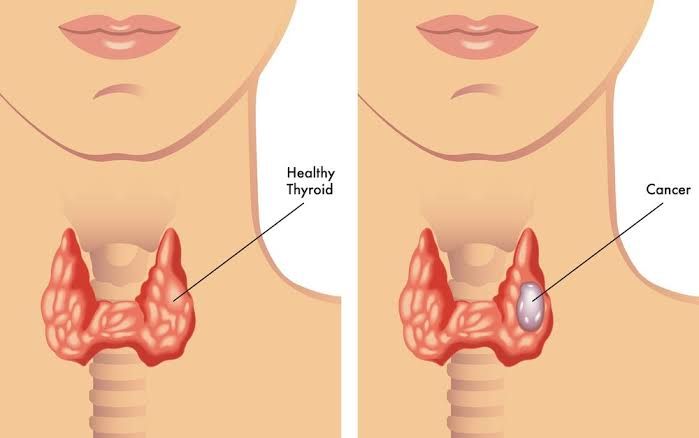
Adenomas and Carcinomas: Differential Diagnosis
Adenomas are benign (non-cancerous) tumors that arise from glandular tissue. In the context of the parathyroid glands, adenomas are the most common cause of primary hyperparathyroidism, accounting for 80–90% of cases. These tumors typically involve one gland and lead to excessive secretion of parathyroid hormone (PTH), causing hypercalcemia. Adenomas are composed mainly of chief cells, which are responsible for PTH production.
Carcinomas, on the other hand, are malignant (cancerous) tumors that can invade surrounding tissues and metastasize to distant organs. Parathyroid carcinoma is extremely rare, representing less than 1% of all cases of primary hyperparathyroidism. It is often difficult to distinguish between parathyroid adenoma and carcinoma based on imaging or laboratory tests alone because both conditions present with elevated calcium levels and PTH secretion. Definitive diagnosis often requires surgical pathology, where features such as capsular invasion, vascular invasion, or metastasis confirm malignancy.
The differential diagnosis between adenoma and carcinoma involves:
- Clinical presentation: Carcinomas may present with more severe symptoms of hypercalcemia (e.g., profound fatigue, bone pain, kidney stones).
- Imaging studies: While imaging can localize abnormal parathyroid glands, it cannot reliably differentiate adenoma from carcinoma.
- Histopathology: Features like mitotic activity, capsular invasion, and adherence to surrounding structures help identify carcinomas.
Types of Malignancies in the Thyroid
Thyroid malignancies include several types:
- Papillary Thyroid Carcinoma (PTC): The most common type (~80% of thyroid cancers). It has an excellent prognosis and often spreads to lymph nodes.
- Follicular Thyroid Carcinoma (FTC): Accounts for ~10–15% of thyroid cancers; it tends to spread hematogenously to bones or lungs.
- Medullary Thyroid Carcinoma (MTC): Arises from parafollicular C cells that produce calcitonin. It may occur sporadically or as part of multiple endocrine neoplasia type 2 (MEN-2).
- Anaplastic Thyroid Carcinoma: A rare but aggressive form with poor prognosis.
- Primary Thyroid Lymphoma: Rarely arises in the thyroid gland; associated with Hashimoto’s thyroiditis.
Primary vs Secondary Hyperparathyroidism
1. Primary Hyperparathyroidism
This condition results from overproduction of PTH due to intrinsic abnormalities in one or more parathyroid glands:
- Causes include a single adenoma (~85%), multiple adenomas (~4–5%), hyperplasia (~10–12%), or rarely carcinoma (<1%).
- Leads to hypercalcemia due to increased bone resorption, renal calcium reabsorption, and intestinal calcium absorption.
2. Secondary Hyperparathyroidism
This occurs as a compensatory response to chronic hypocalcemia caused by external factors such as:
- Chronic kidney disease (CKD): Reduced vitamin D activation leads to hypocalcemia.
- Vitamin D deficiency or malabsorption.
- Chronic illnesses causing low calcium levels.
Unlike primary hyperparathyroidism, secondary hyperparathyroidism typically presents with normal or low serum calcium levels but elevated PTH.
Parathyroid Hyperplasia vs Parathyroid Adenoma
1. Parathyroid Hyperplasia
- Involves enlargement of all four parathyroid glands.
- Often sporadic but may be associated with genetic syndromes like MEN-1 or MEN-2A.
- Causes primary hyperparathyroidism in ~5–15% of cases.
- Treatment involves removal of 3½ glands.
2. Parathyroid Adenoma
- Typically affects only one gland.
- Responsible for ~80–90% of primary hyperparathyroidism cases.
- Surgical removal cures the condition in most patients.
Key difference: Hyperplasia affects all four glands symmetrically while adenomas usually involve a single gland.
Hypoparathyroidism: Clinical Manifestations and Etiology
Clinical Manifestations
Hypoparathyroidism results in hypocalcemia due to insufficient PTH secretion:
- Neuromuscular symptoms: Tetany, muscle cramps, paresthesia (tingling around lips/fingers).
- Severe cases: Seizures or laryngospasm.
- Chvostek’s sign (facial twitching when tapping facial nerve) and Trousseau’s sign (carpal spasm after inflating a blood pressure cuff) are classic findings.
Etiology
The most common causes include:
- Post-surgical damage/removal during thyroidectomy or neck surgery (~0.5–6.6% incidence).
- Autoimmune destruction as part of polyglandular autoimmune syndromes.
- Genetic disorders like DiGeorge syndrome or pseudohypoparathyroidism (tissue resistance to PTH despite normal/elevated levels).
- Other rare causes include magnesium deficiency or infiltrative diseases affecting the parathyroids.


