Structure of Calcitonin
Calcitonin is a peptide hormone composed of 32 amino acids. Its structure includes a disulfide bridge between cysteine residues at positions 1 and 7, which stabilizes the molecule. The carboxy-terminal end contains proline, which is essential for its biological activity. The sequence of calcitonin varies slightly among species, but the human calcitonin sequence is distinct from that of other animals like salmon, which has been used therapeutically due to its higher potency.
Major Physiological Actions of Calcitonin
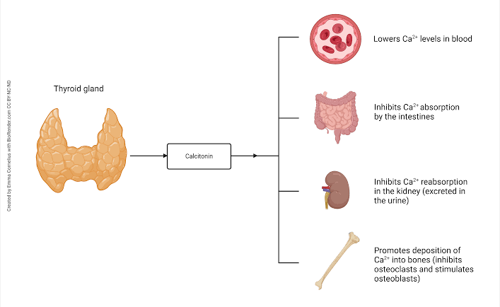
- Reduction in Blood Calcium Levels:
Calcitonin primarily acts to lower blood calcium levels when they are elevated (hypercalcemia). It achieves this by:- Inhibiting osteoclast activity in bones, thereby reducing bone resorption and calcium release into the bloodstream.
- Promoting calcium deposition into bone by facilitating osteoblast activity.
- Inhibition of Bone Resorption:
Calcitonin directly suppresses the breakdown of bone matrix by osteoclasts, leading to decreased release of calcium and phosphate from bones.
- Renal Effects:
It reduces renal tubular reabsorption of calcium and phosphate, promoting their excretion in urine.
- Role in Vitamin D Regulation:
Calcitonin stimulates the production of 1,25-dihydroxyvitamin D (active vitamin D) in the kidney through mechanisms distinct from parathyroid hormone (PTH).
- Potential Role During Pregnancy and Lactation:
During pregnancy and lactation, calcitonin levels increase along with vitamin D levels to facilitate maternal calcium transfer to the fetus or infant while protecting maternal bone health.
- Therapeutic Applications:
Due to its ability to inhibit bone resorption, calcitonin has been used clinically for conditions such as osteoporosis, Paget’s disease, and hypercalcemia.
Regulation of Calcitonin Secretion
The secretion of calcitonin is tightly regulated by several factors:
- Serum Calcium Levels:
- High serum calcium levels stimulate calcitonin secretion via activation of calcium-sensing receptors (CaSR) on thyroid C-cells.
- Conversely, low serum calcium levels suppress calcitonin secretion.
- Gastrointestinal Hormones:
- Hormones like gastrin can stimulate calcitonin release postprandially (after eating), suggesting a potential role in depositing dietary calcium into bones.
- Vitamin D Feedback Mechanism:
- Active vitamin D (1,25-dihydroxyvitamin D) suppresses calcitonin gene transcription as part of a feedback loop.
- Adaptation to Chronic Conditions:
- Chronic hypercalcemia can lead to depletion or adaptation in calcitonin secretion over time.
- In contrast, chronic hypocalcemia may enhance thyroidal calcitonin content and responsiveness.
- Other Factors Influencing Secretion:
- Age and gender differences have been observed; women tend to have lower baseline levels than men.
- Certain pathological conditions like medullary thyroid carcinoma can cause excessive secretion.
Comparison Between PTH and Calcitonin as Regulators of Calcium Levels
Parathyroid Hormone (PTH) and calcitonin are two key hormones that regulate calcium levels in the body, but they function in opposite ways to maintain calcium homeostasis. Below is a detailed comparison of their roles, mechanisms, and physiological significance:
1. Source of Hormones
- PTH: Parathyroid hormone is secreted by the parathyroid glands, which are four small glands located behind the thyroid gland.
- Calcitonin: Calcitonin is produced by parafollicular cells (C-cells) in the thyroid gland.
2. Primary Function
- PTH: The primary role of PTH is to increase blood calcium levels when they fall below normal. It achieves this through several mechanisms:
- Stimulating bone resorption by activating osteoclasts, which break down bone tissue to release calcium into the bloodstream.
- Enhancing calcium reabsorption in the kidneys, reducing urinary calcium excretion.
- Promoting the activation of vitamin D (calcitriol) in the kidneys, which increases intestinal absorption of dietary calcium.
- Calcitonin: Calcitonin works to decrease blood calcium levels when they rise above normal. It does so by:
- Inhibiting osteoclast activity, thereby reducing bone resorption and promoting calcium deposition into bones.
- Decreasing renal reabsorption of calcium, leading to increased excretion of calcium in urine.
3. Mechanism of Action
- PTH:
- PTH binds to receptors on osteoblasts (bone-forming cells), stimulating them to produce RANKL (Receptor Activator for Nuclear Factor κ B Ligand). RANKL activates osteoclasts, which break down bone matrix and release stored calcium into the bloodstream.
- In the kidneys, PTH upregulates specific transport proteins in the distal convoluted tubule to enhance calcium reabsorption while simultaneously promoting phosphate excretion.
- PTH indirectly increases intestinal absorption of calcium by stimulating the production of calcitriol (active vitamin D).
- Calcitonin:
- Calcitonin directly inhibits osteoclast activity, preventing excessive breakdown of bone tissue and reducing the release of stored calcium into circulation.
- It also reduces renal tubular reabsorption of calcium, increasing urinary excretion.
4. Regulation
- Both hormones are regulated by blood calcium levels through negative feedback loops:
- When blood calcium levels drop below normal, PTH secretion increases; when levels normalize or rise too high, PTH secretion decreases.
- Conversely, when blood calcium levels rise above normal, calcitonin secretion increases; as levels normalize or drop too low, calcitonin secretion decreases.
5. Relative Importance
- PTH: Plays a dominant role in maintaining long-term blood calcium homeostasis. Its effects on bones, kidneys, and intestines ensure that blood calcium remains within a narrow range essential for vital physiological functions like muscle contraction and neurotransmission.
- Calcitonin: While it contributes to lowering elevated blood calcium levels temporarily, its role is less critical compared to PTH. Studies suggest that calcitonin’s absence does not significantly disrupt overall calcium balance because other mechanisms can compensate for its function.
6. Clinical Relevance
- Dysregulation of these hormones can lead to significant health issues:
- Excessive PTH secretion (e.g., hyperparathyroidism) causes hypercalcemia (high blood calcium), leading to symptoms such as kidney stones, osteoporosis, fatigue, and cardiac arrhythmias.
- Insufficient PTH secretion (e.g., hypoparathyroidism) results in hypocalcemia (low blood calcium), causing muscle spasms, seizures, and cardiac issues due to increased neuromuscular excitability.
- Abnormal calcitonin levels are generally asymptomatic but may indicate medullary thyroid cancer or C-cell hyperplasia if excessively high.


