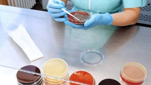
Selection, Collection, and Transportation of Sputum Sample
Selection
Sputum samples are critical for diagnosing respiratory infections such as tuberculosis (TB). The sample must be collected from deep within the lungs to ensure it contains mucus rather than saliva. This is essential because saliva contamination can dilute or obscure the presence of pathogens like Mycobacterium tuberculosis.
Collection
- Preparation:
- The patient should drink plenty of fluids (e.g., water or tea) the night before to loosen mucus in the lungs.
- Collection is ideally done early in the morning when bacterial concentration in sputum is highest.
- Patients should avoid eating or drinking anything before collecting the sample.
- Procedure:
- Brush teeth and rinse the mouth with water (without antiseptic mouthwash) to reduce contamination.
- Take deep breaths to loosen mucus in the lungs.
- Cough deeply to bring up sputum from the lower respiratory tract.
- Spit the sputum into a sterile container provided by healthcare professionals until it reaches approximately 1 teaspoon (5 mL).
- Alternative Methods:
- If patients cannot produce sputum naturally, steam inhalation or hot showers may help induce coughing.
- For severe cases where natural collection fails, bronchoscopy may be performed to retrieve sputum directly from the lungs.
Transportation
- The collected sample must be transported promptly to a laboratory for analysis.
- If immediate transport is not possible:
- Refrigerate the sample at 4°C for up to 24 hours.
- Avoid freezing or storing at room temperature as this can degrade bacterial viability.
Cultivation of Acid-Fast and Non-Acid-Fast Bacteria
Acid-Fast Bacteria Cultivation
Acid-fast bacteria like Mycobacterium tuberculosis are cultivated using specialized media due to their slow growth and unique cell wall properties.
- Lowenstein-Jensen (LJ) Medium:
- LJ medium is an egg-based solid medium enriched with malachite green dye that inhibits contaminants while promoting mycobacterial growth.
- It supports visible colony formation over weeks due to M. tuberculosis’s slow doubling time (~18–24 hours).
- Incubation Conditions:
- Samples are incubated at 35–37°C in an environment with adequate humidity.
- Growth typically appears after 2–8 weeks depending on bacterial load.
Non-Acid-Fast Bacteria Cultivation
Non-acid-fast bacteria (e.g., Staphylococcus aureus) grow more rapidly on general-purpose media such as nutrient agar or blood agar.
- Incubation conditions vary but are generally faster than acid-fast organisms (growth within 24–48 hours).
- These bacteria lack mycolic acids in their cell walls, making them easier to stain and cultivate.
Ziehl-Neelsen Staining Procedure
The Ziehl-Neelsen stain is a differential staining technique used specifically for identifying acid-fast bacilli like Mycobacterium tuberculosis.
Steps:
- Preparation:
- Prepare a smear by spreading a small amount of sputum onto a clean glass slide using an inoculating loop.
- Allow it to air dry completely before heat-fixing by passing it through a flame.
- Staining Process:
- Apply carbol fuchsin dye over the smear and heat gently until steaming without boiling (this helps penetrate waxy mycolic acids in acid-fast bacteria).
- Rinse with water after cooling.
- Decolorization:
- Wash with an acid-alcohol solution (e.g., 3% hydrochloric acid in ethanol). Acid-fast organisms retain carbol fuchsin due to their lipid-rich cell walls.
- Counterstaining:
- Apply methylene blue or malachite green as a counterstain for non-acid-fast cells.
- Rinse again with water and allow drying.
Visualization and Observation Under Microscope
After Ziehl-Neelsen staining:
- Use bright-field microscopy at high magnification (1000x) with oil immersion for observation.
- Acid-fast bacilli appear as bright red rods against a blue or green background depending on counterstain used.
- Non-acid-fast organisms will take up only the counterstain color.
Familiarity with Lowenstein-Jensen Medium
Lowenstein-Jensen medium plays an essential role in culturing Mycobacterium species:
- Composition includes eggs, glycerol, potato flour, and malachite green dye which suppresses contaminant growth while allowing slow-growing mycobacteria colonies.
- Colonies appear rough, buff-colored (“cauliflower-like”) after several weeks of incubation.
Preparation of Slides from Sputum for Staining
To prepare slides:
- Place a small drop of fresh sputum on one end of a clean glass slide using an inoculating loop.
- Spread evenly across the slide surface into a thin layer using circular motions.
- Air-dry completely before heat-fixing by passing through flame three times briefly.
- Proceed with Ziehl-Neelsen staining as outlined above.


