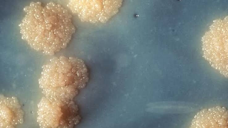
Morphology, Structure, Staining, and Cultural Characteristics of Mycobacterium tuberculosis
Mycobacterium tuberculosis (M. tuberculosis) is a rod-shaped, non-motile, non-spore-forming bacterium that belongs to the family Mycobacteriaceae. It measures approximately 1–4 µm in length and 0.2–0.6 µm in width. The organism is an obligate aerobe and grows best in tissues with high oxygen concentrations, such as the lungs.
Structurally, M. tuberculosis has a unique cell wall rich in lipids, including mycolic acids, glycolipids, and waxes. This lipid-rich cell wall is responsible for its acid-fast property and contributes significantly to its virulence by providing resistance to desiccation, disinfectants, and antibiotics.
- Staining Characteristics: M. tuberculosis is classified as an acid-fast bacillus (AFB). It does not stain well with Gram staining due to its lipid-rich cell wall but retains carbol fuchsin dye when treated with acid-alcohol during Ziehl-Neelsen or Kinyoun staining methods. This acid-fastness is a hallmark of the genus Mycobacterium.
- Cultural Characteristics: M. tuberculosis grows slowly on artificial media due to its long generation time (15–20 hours). It requires enriched media like Löwenstein-Jensen medium or Middlebrook 7H10/7H11 agar for optimal growth. Colonies appear rough, buff-colored (“breadcrumb-like”), and dry after 2–6 weeks of incubation at 35–37°C.
Relation of Structure to Virulence and Pathogenesis
The structural components of M. tuberculosis play critical roles in its virulence and ability to cause disease:
- Lipid-Rich Cell Wall: The mycolic acids and other lipids in the cell wall provide resistance against host immune responses (e.g., phagocytosis) and environmental stressors like antibiotics.
- Cord Factor (Trehalose Dimycolate): This glycolipid inhibits macrophage activation and induces granuloma formation by disrupting mitochondrial function in host cells.
- ESX Secretion Systems: Proteins secreted via ESX systems (e.g., ESAT-6) modulate host immunity by interfering with antigen presentation pathways.
- Lipoarabinomannan (LAM): LAM suppresses T-cell activation by inhibiting interferon-gamma production while promoting survival within macrophages.
These structural features enable M. tuberculosis to evade immune defenses, establish latent infections within granulomas, and reactivate under favorable conditions.
Range of Pathogenicity
M. tuberculosis primarily causes pulmonary TB but can also lead to extrapulmonary TB affecting lymph nodes, bones (Pott’s disease), meninges (TB meningitis), kidneys, or skin.
- Latent Tuberculosis Infection (LTBI): In most individuals (~90%), the immune system contains the infection without symptoms.
- Active Tuberculosis Disease (ATB): In ~5–10% of infected individuals or those with weakened immunity (e.g., HIV/AIDS), LTBI progresses into active disease characterized by cough, fever, weight loss, hemoptysis, and night sweats.
Resistance
M. tuberculosis exhibits intrinsic resistance due to its impermeable cell wall but can acquire drug resistance through mutations:
- Multidrug-Resistant TB (MDR-TB): Resistance to rifampin (RIF) and isoniazid (INH).
- Extensively Drug-Resistant TB (XDR-TB): Resistance to RIF/INH plus fluoroquinolones and second-line injectable drugs.
Antigenic Structure
Key antigens include:
- Early Secretory Antigen Target-6 (ESAT-6)
- Culture Filtrate Protein-10 (CFP-10)
These antigens are used diagnostically in interferon-gamma release assays (IGRAs) for detecting latent infections.
Virulence Mechanisms
M. tuberculosis employs multiple mechanisms:
- Survival within macrophages by inhibiting phagosome maturation.
- Induction of granuloma formation for persistence.
- Modulation of host immune responses via secreted proteins like ESAT-6.
Antimicrobial Susceptibility
First-line drugs include:
- Rifampin
- Isoniazid
- Pyrazinamide
- Ethambutol
Second-line drugs are used for MDR/XDR-TB cases.
Tuberculosis: Routes of Infection & Reactivation
TB spreads via inhalation of aerosolized droplets containing Mtb from an infected individual’s cough or sneeze.
Reactivation occurs when latent bacteria become active due to immunosuppression caused by factors like HIV/AIDS or malnutrition.
Immunity, Transmission & Epidemiology
- Immunity:
- Cell-mediated immunity plays a central role; CD4+ T-cells produce interferon-gamma to activate macrophages.
- Granulomas form around infected macrophages as a containment strategy.
- Transmission:
- Airborne transmission via respiratory droplets.
- Close contact increases risk.
- Epidemiology:
- One-quarter of the global population has LTBI.
- High prevalence in low-income regions with poor healthcare access.
Laboratory Diagnosis
- Microscopy:
- Acid-fast staining using Ziehl-Neelsen or fluorescent auramine-rhodamine stains.
- Culture:
- Löwenstein-Jensen medium or liquid culture systems like MGIT for faster results.
- Molecular Tests:
- GeneXpert MTB/RIF detects Mtb DNA and rifampin resistance within hours.
- Immunologic Tests:
- Tuberculin Skin Test (TST).
- Interferon-Gamma Release Assays (IGRAs).
- Radiographic Imaging:
- Chest X-rays reveal cavitations or infiltrates typical of pulmonary TB.
Anti-Tuberculosis Drugs and Multidrug-Resistant Organisms
Tuberculosis (TB), caused by Mycobacterium tuberculosis (Mtb), is treated using a combination of first-line and second-line drugs. The standard treatment for drug-sensitive TB involves a six-month regimen of multiple antibiotics, while multidrug-resistant TB (MDR-TB) requires more complex and prolonged therapies.
First-Line Anti-Tuberculosis Drugs
The first-line drugs are the most effective and least toxic options for treating TB. These include:
- Isoniazid (INH): Inhibits mycolic acid synthesis, which is essential for the bacterial cell wall.
- Rifampicin (RIF): Inhibits bacterial RNA polymerase, preventing transcription.
- Pyrazinamide (PZA): Disrupts membrane transport and energy production in acidic environments.
- Ethambutol (EMB): Inhibits arabinosyl transferase enzymes, disrupting cell wall synthesis.
These drugs are used in combination to prevent resistance development and ensure complete eradication of the bacteria.
Second-Line Anti-Tuberculosis Drugs
Second-line drugs are used when resistance to first-line agents occurs or when patients cannot tolerate them. These include:
- Fluoroquinolones: Such as levofloxacin or moxifloxacin, which inhibit DNA gyrase.
- Injectable Agents: Such as amikacin, kanamycin, or capreomycin.
- Newer Agents: Bedaquiline and delamanid target ATP synthase and mycolic acid biosynthesis, respectively.
Multidrug-Resistant Tuberculosis (MDR-TB)
MDR-TB refers to strains of Mtb resistant to at least isoniazid and rifampicin, the two most potent first-line drugs. Extensively drug-resistant TB (XDR-TB) includes additional resistance to fluoroquinolones and at least one injectable second-line agent.
Immunoprophylaxis: Definition and Vaccines Used
Definition of Immunoprophylaxis
Immunoprophylaxis refers to the prevention of disease through vaccination or passive immunization strategies that stimulate or provide immunity against specific pathogens.
Vaccines Used Against Tuberculosis
The primary vaccine currently available for TB is the Bacille Calmette–Guérin (BCG) vaccine. However, its efficacy varies geographically due to factors such as environmental mycobacteria exposure and genetic differences among populations.
Strategies of Vaccination
- Prophylactic Vaccination:
- The BCG vaccine is administered primarily to infants in high-burden countries shortly after birth.
- It provides protection against severe forms of TB such as meningitis and miliary TB but has variable efficacy against pulmonary TB.
- Therapeutic Vaccination:
- Designed as adjunctive therapy for active TB or prevention of relapse after treatment completion.
- Examples include Mycobacterium vaccae-based vaccines, RUTI (liposomal fragments of Mtb), and other candidates currently under clinical trials.
Role of PPD Testing and Its Significance
The Purified Protein Derivative (PPD) test, also known as the Mantoux tuberculin skin test, is a diagnostic tool used to detect latent tuberculosis infection (LTBI). It involves intradermal injection of PPD tuberculin into the forearm skin.
Mechanism
- PPD contains antigens derived from Mycobacterium tuberculosis.
- If an individual has been exposed to Mtb, their immune system will recognize these antigens, leading to a delayed-type hypersensitivity reaction mediated by T-cells within 48–72 hours.
Interpretation
- A raised induration at the injection site indicates prior exposure to Mtb or vaccination with BCG.
- The size threshold for a positive result depends on risk factors:
- ≥5 mm: Positive in high-risk individuals such as HIV-positive patients.
- ≥10 mm: Positive in moderate-risk groups like healthcare workers.
- ≥15 mm: Positive in individuals with no known risk factors.
Significance
- Identifies individuals with LTBI who may benefit from preventive therapy.
- Helps control TB transmission by targeting latent infections before they progress to active disease.
In summary, anti-tuberculosis drugs include both first-line agents like isoniazid and rifampicin, as well as second-line options for MDR-TB cases such as fluoroquinolones and bedaquiline. Immunoprophylaxis involves vaccines like BCG for prevention; therapeutic vaccines are being developed for adjunctive treatment strategies. The PPD test remains critical for diagnosing latent infections and guiding preventive measures.


