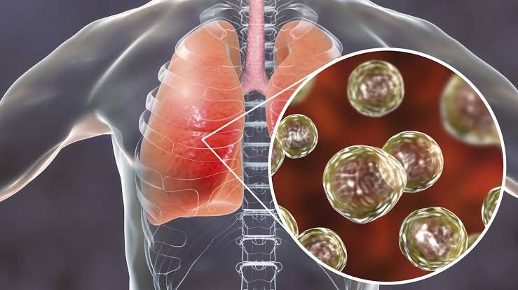
Structure, Clinical Classification, and Significance in Disease Process
Fungi involved in respiratory tract infections are classified into two main categories: endemic fungi and opportunistic fungi. Their structure and clinical classification play a significant role in their pathogenicity.
1. Endemic Fungi
These fungi can cause disease in both immunocompetent and immunocompromised individuals. They are geographically restricted to specific regions.
- Histoplasma capsulatum: A dimorphic fungus that exists as a mold in the environment and as a yeast at body temperature. It is found in soil enriched with bird or bat droppings. Histoplasmosis can range from asymptomatic infection to severe disseminated disease.
- Coccidioides immitis/posadasii: Another dimorphic fungus that exists as mold in the environment and forms spherules containing endospores in human tissue. It causes coccidioidomycosis, also known as “Valley Fever,” which may present as mild respiratory illness or disseminated disease.
- Blastomyces dermatitidis: This dimorphic fungus is found in moist soil and decaying organic matter. It causes blastomycosis, which can involve the lungs, skin, bones, and other organs.
- Paracoccidioides brasiliensis: Found primarily in Central and South America, it causes paracoccidioidomycosis with pulmonary involvement often progressing to systemic disease.
2. Opportunistic Fungi
These fungi primarily infect immunocompromised individuals.
- Aspergillus species (e.g., Aspergillus fumigatus): A ubiquitous mold that produces airborne conidia (spores). It causes invasive aspergillosis, allergic bronchopulmonary aspergillosis (ABPA), or aspergilloma (fungal ball).
- Cryptococcus neoformans/gattii: Encapsulated yeasts found in soil contaminated with bird droppings. Cryptococcus neoformans primarily affects immunocompromised patients (e.g., those with AIDS), causing cryptococcal meningitis after pulmonary infection.
- Pneumocystis jirovecii: An atypical fungus causing Pneumocystis pneumonia (PCP) predominantly in HIV/AIDS patients or those on prolonged corticosteroid therapy.
- Candida species (e.g., Candida albicans): Although part of normal flora, Candida can cause pneumonia under rare circumstances, particularly in severely immunosuppressed hosts.
- Mucorales species (e.g., Rhizopus spp.): These molds cause mucormycosis, an aggressive infection seen mainly in diabetic ketoacidosis or neutropenic patients.
Epidemiology
The prevalence of fungal infections varies based on geographic distribution and host immune status:
- Endemic mycoses like histoplasmosis are common along river valleys such as the Ohio and Mississippi River Valleys.
- Coccidioidomycosis is endemic to arid regions of the southwestern United States.
- Opportunistic fungal infections like invasive aspergillosis occur globally but are more frequent among immunosuppressed populations.
Pathogenesis
Fungal pathogens initiate infection by inhalation of spores or conidia into the respiratory tract:
- In healthy individuals:
- The innate immune system typically eliminates inhaled spores via alveolar macrophages and neutrophils.
- Adaptive immunity involving Th1 cells producing interferon-gamma plays a critical role.
- In immunocompromised individuals:
- Impaired phagocytosis allows fungal spores to germinate into hyphae (in molds) or yeast forms.
- Dissemination occurs via hematogenous spread to extrapulmonary sites such as the brain, liver, spleen, or skin.
Clinical Presentation
The clinical manifestations depend on the type of fungus and host immune status:
- Endemic Mycoses:
- Acute pulmonary symptoms include fever, cough, chest pain, dyspnea.
- Chronic cases may lead to cavitary lesions resembling tuberculosis.
- Opportunistic Mycoses:
- Aspergillosis presents with hemoptysis or invasive disease leading to necrotizing pneumonia.
- Cryptococcal infections often progress from asymptomatic lung colonization to meningitis.
- Pneumocystis pneumonia presents with progressive dyspnea, hypoxia, fever without significant radiographic findings initially.
Laboratory Diagnosis
Diagnosis involves a combination of clinical suspicion, imaging studies (e.g., CT scans), microbiological tests:
- Direct Microscopy:
- Stains like KOH preparation for fungal elements; India ink for Cryptococcus capsule visualization.
- Culture:
- Growth on Sabouraud dextrose agar confirms fungal identification but may take weeks for some species.
- Antigen Detection:
- Galactomannan assay for Aspergillus; cryptococcal antigen test for Cryptococcus.
- Molecular Methods:
- PCR-based assays provide rapid identification of fungal DNA.
- Imaging Studies:
- Chest X-rays/CT scans reveal nodules, cavitations typical of fungal pneumonias.
Treatment and Antifungal Drugs
Treatment depends on the causative organism:
- Polyenes:
- Amphotericin B binds ergosterol disrupting fungal cell membranes.
- Toxicity includes nephrotoxicity; liposomal formulations reduce side effects.
- Azoles:
- Fluconazole inhibits ergosterol synthesis; used for Cryptococcus and endemic mycoses.
- Itraconazole is effective against histoplasmosis/blastomycosis but has hepatotoxicity risks.
- Echinocandins:
- Caspofungin inhibits β-glucan synthesis affecting fungal cell walls; used for Candida/Aspergillus infections.
- Trimethoprim-Sulfamethoxazole:
- First-line treatment for Pneumocystis jirovecii pneumonia (PCP).
Preventive Measures
Preventive strategies include:
- Avoiding exposure to high-risk environments such as areas rich in bird/bat droppings for endemic mycoses.
- Prophylactic antifungals like posaconazole for high-risk neutropenic patients reduce invasive fungal infections.
- Vaccination research is ongoing but not yet available clinically.
Role of Immune System
The immune response involves both innate mechanisms (macrophages/neutrophils) and adaptive immunity (Th1/Th17 cells). Immunosuppression due to HIV/AIDS or chemotherapy significantly increases susceptibility to severe fungal diseases by impairing these defenses.


