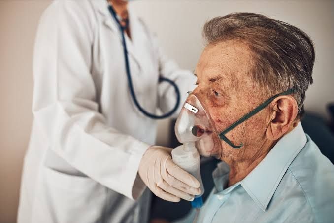
Definition of Restrictive Lung Disease
Restrictive lung disease refers to a group of respiratory disorders characterized by reduced lung expansion and diminished total lung capacity (TLC). This condition can result from intrinsic factors, such as diseases affecting the lung parenchyma (e.g., interstitial lung diseases), or extrinsic factors, such as abnormalities in the pleura, chest wall, or neuromuscular system. The hallmark feature is difficulty in fully inflating the lungs with air due to stiffness or restriction in the lungs or surrounding structures.
Principles, Causes, and Mechanisms in Acute Restrictive Lung Disease: Acute Respiratory Distress Syndrome (ARDS)
Acute Respiratory Distress Syndrome (ARDS) in Adults
ARDS is a severe form of acute restrictive lung disease caused by diffuse alveolar damage leading to non-cardiogenic pulmonary edema. It is characterized by rapid onset of hypoxemia and bilateral infiltrates on imaging without evidence of left atrial hypertension.
Mechanisms and Pathophysiology:
- Injury to Alveolar-Capillary Barrier: ARDS begins with direct (e.g., pneumonia, aspiration) or indirect (e.g., sepsis, trauma) injury to the alveolar-capillary barrier.
- Inflammation and Increased Permeability: Pro-inflammatory cytokines like IL-6 and TNF-α are released, increasing vascular permeability. This leads to leakage of protein-rich fluid into the alveoli.
- Impaired Gas Exchange: Fluid accumulation disrupts surfactant production, causing alveolar collapse (atelectasis), decreased compliance, and impaired oxygenation.
- Fibroproliferative Phase: Persistent inflammation may lead to fibrosis during later stages.
Neonatal ARDS (Hyaline Membrane Disease)
In newborns, ARDS often results from surfactant deficiency due to immature lungs:
- Premature infants lack sufficient surfactant production by type II pneumocytes.
- Surfactant deficiency increases surface tension within alveoli, causing widespread atelectasis.
- Hypoxemia ensues due to ventilation-perfusion mismatch.
Both adult and neonatal ARDS require supportive care such as mechanical ventilation and oxygen therapy.
Pathology of Idiopathic Pulmonary Fibrosis
Idiopathic pulmonary fibrosis (IPF) is a chronic progressive interstitial lung disease characterized by scarring (fibrosis) of unknown cause. It falls under the category of restrictive lung diseases.
Pathological Features:
- Usual Interstitial Pneumonia (UIP): The hallmark histopathological pattern seen in IPF includes:
- Patchy areas of fibrosis alternating with normal lung tissue.
- Subpleural and basal predominance.
- Temporal heterogeneity: coexistence of early fibroblastic foci with dense collagen deposition.
- Honeycombing: cystic spaces lined by bronchiolar epithelium.
- Mechanism:
- Repeated micro-injuries to alveolar epithelial cells trigger abnormal wound healing responses.
- Dysregulated fibroblast activity leads to excessive extracellular matrix deposition and scarring.
The disease progresses over time with worsening dyspnea and reduced gas exchange efficiency.
Common Causes of Pulmonary Fibrosis with Emphasis on Sarcoidosis Pathology
Pulmonary fibrosis can result from various conditions:
Common Causes:
- Idiopathic pulmonary fibrosis (IPF).
- Connective tissue diseases like systemic sclerosis or rheumatoid arthritis.
- Environmental/occupational exposures such as asbestos or silica dust.
- Drug-induced fibrosis from medications like amiodarone or methotrexate.
- Radiation-induced damage following cancer treatment.
Sarcoidosis Pathology:
Sarcoidosis is an inflammatory disease that frequently affects the lungs (~90% cases). It is characterized by granuloma formation—organized clusters of macrophages surrounded by lymphocytes—and can progress to pulmonary fibrosis in chronic cases.
Key Features:
- Non-caseating granulomas are the pathological hallmark.
- Granulomas are composed of epithelioid macrophages surrounded by T-helper lymphocytes.
- Chronic inflammation can lead to scarring and irreversible fibrotic changes in advanced stages.
Mechanism:
The exact cause remains unknown but involves an exaggerated immune response triggered by environmental antigens in genetically predisposed individuals.
Clinical Implications:
While many cases resolve spontaneously, some progress to chronic sarcoidosis-associated pulmonary fibrosis, which may require corticosteroids or immunosuppressants for management.
Causes and Pathology of Pneumoconiosis
Pneumoconiosis refers to a group of occupational lung diseases caused by the inhalation of mineral dusts, which leads to chronic inflammation and fibrosis in the lungs. The primary causes include prolonged exposure to specific types of dust, such as coal dust, silica, and asbestos. These diseases are classified under interstitial lung diseases (ILDs) and are characterized by scarring (fibrosis) in the lung interstitium.
Causes
- Coal Workers’ Pneumoconiosis (CWP): Caused by inhalation of coal dust over many years. It can progress from a simple form (often asymptomatic) to complicated pneumoconiosis or “progressive massive fibrosis” (PMF), which involves severe scarring and respiratory dysfunction.
- Silicosis: Results from inhaling crystalline silica dust, commonly found in occupations like mining, stone cutting, masonry, and ceramics. Silica particles trigger inflammation and fibrosis in the lungs.
- Asbestosis: Caused by inhalation of asbestos fibers, typically found in construction, shipbuilding, and demolition work. Asbestos exposure can also lead to mesothelioma.
Pathology
- Inhaled mineral dust particles deposit in the alveoli.
- Macrophages attempt to engulf these particles but fail due to their size or chemical properties.
- This triggers an inflammatory response with cytokine release that promotes fibroblast proliferation.
- Over time, this leads to collagen deposition and fibrosis within the lung interstitium.
- Advanced cases may result in “honeycombing,” where cystic spaces develop within fibrotic tissue.
Causes and Pathology of Asbestosis and Mesothelioma
Asbestosis
Causes:
- Prolonged inhalation of asbestos fibers (chrysotile, amosite, crocidolite).
- Common sources include insulation materials, roofing products, brake pads, and demolition work involving older buildings containing asbestos.
Pathology:
- Asbestos fibers lodge in the alveolar ducts and peribronchiolar regions.
- Alveolar macrophages engulf these fibers but cannot degrade them effectively.
- Persistent inflammation leads to fibroblast activation and collagen deposition.
- Fibrosis predominantly affects lower lung fields.
- Severe cases show honeycombing on imaging studies.
Mesothelioma
Causes:
- Almost exclusively linked to asbestos exposure; even brief exposures can increase risk significantly.
- There is often a latency period of 20–40 years between exposure and disease onset.
Pathology:
- Mesothelioma arises from mesothelial cells lining the pleura (or peritoneum).
- Asbestos fibers cause direct damage to DNA or induce oxidative stress leading to genetic mutations.
- Tumors form along pleural surfaces causing thickening, effusions, chest pain, dyspnea, and eventually respiratory failure.
Pulmonary Hemorrhage Syndromes Leading to Pulmonary Fibrosis
Pulmonary hemorrhage syndromes involve bleeding into the alveoli or lung parenchyma. Chronic or recurrent episodes can lead to pulmonary fibrosis due to repeated cycles of injury and repair.
Examples
- Goodpasture’s Syndrome
- A rare autoimmune disorder caused by anti-glomerular basement membrane antibodies targeting both renal glomeruli and alveolar capillaries.
- Symptoms: Hemoptysis (coughing up blood), hematuria (blood in urine), anemia.
- Pathology: Recurrent alveolar hemorrhage leads to hemosiderin-laden macrophages accumulating in the lungs with subsequent fibrosis.
- Idiopathic Pulmonary Hemosiderosis
- A rare condition seen primarily in children under seven years old.
- Symptoms: Coughing up blood (hemoptysis), dyspnea (shortness of breath).
- Pathology: Repeated episodes of alveolar hemorrhage result in iron deposition within lung tissue followed by fibrosis.
- Systemic Vasculitides
- Diseases like granulomatosis with polyangiitis (formerly Wegener’s granulomatosis) can cause pulmonary hemorrhage as part of systemic small-vessel vasculitis.
- Pathology: Necrotizing granulomas damage capillaries leading to bleeding; chronic inflammation promotes fibrotic changes over time.
- Lupus Erythematosus
- Systemic lupus erythematosus may involve diffuse alveolar hemorrhage as part of its pulmonary manifestations.
- Chronic episodes contribute to scarring within lung tissues.


