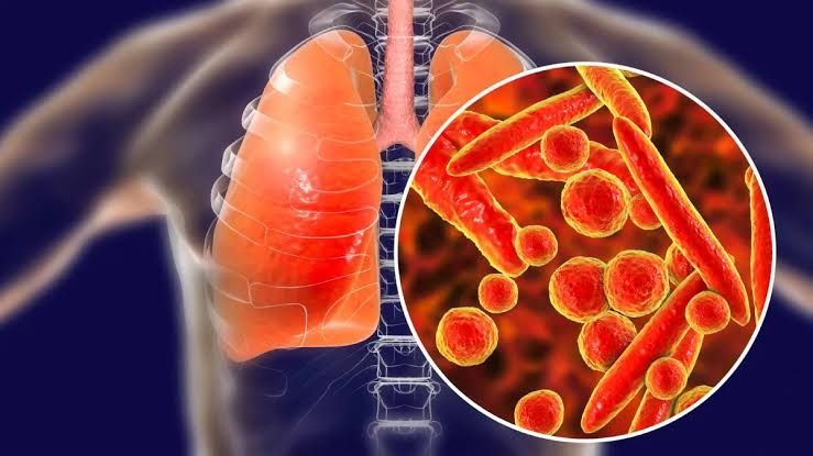
Structure and Morphology
Mycoplasma pneumoniae:
- Mycoplasma species are unique among bacteria because they lack a cell wall. This absence of a rigid peptidoglycan layer makes them pleomorphic (variable in shape) and resistant to antibiotics that target cell wall synthesis, such as beta-lactams.
- They are the smallest free-living organisms, with a size of approximately 0.2–0.3 µm, allowing them to pass through filters that typically retain bacteria.
- The membrane contains sterols, which are acquired from the host and provide structural stability.
- Virulence factors include adhesins like P1 protein, which facilitates attachment to respiratory epithelial cells, leading to localized damage.
Legionella pneumophila:
- Legionella species are gram-negative bacilli but often appear pleomorphic under certain conditions. They require specialized media containing L-cysteine and iron for growth.
- The outer membrane contains lipopolysaccharides (LPS), which contribute to immune evasion. Additionally, pili and flagella enhance adherence and motility.
- Intracellular replication within macrophages is a hallmark of its pathogenesis. This is facilitated by the Dot/Icm type IV secretion system, which injects effector proteins into host cells to manipulate cellular processes.
Virulence and Pathogenesis
Mycoplasma pneumoniae:
- Pathogenesis begins with adhesion to ciliated epithelial cells in the respiratory tract via P1 adhesin. This disrupts ciliary function (ciliostasis), impairing mucociliary clearance.
- The organism produces hydrogen peroxide and superoxide radicals, causing oxidative damage to host tissues.
- Immune-mediated mechanisms also play a role in tissue injury. For example, cross-reactivity between bacterial antigens and host tissues can lead to extrapulmonary manifestations such as hemolytic anemia or Stevens-Johnson syndrome.
Legionella pneumophila:
- After inhalation of aerosolized contaminated water droplets, Legionella reaches the alveoli where it is phagocytosed by alveolar macrophages.
- Instead of being destroyed, Legionella manipulates the phagosome using its Dot/Icm secretion system to create a replicative vacuole that avoids lysosomal fusion.
- Intracellular replication leads to cell lysis and release of bacteria into surrounding tissues.
- The immune response involves both innate mechanisms (macrophage activation via cytokines like interferon-gamma) and adaptive immunity (T-cell-mediated responses). However, immunocompromised individuals have impaired defenses against this pathogen.
Clinical Presentation
Mycoplasma pneumoniae:
- Causes atypical pneumonia (“walking pneumonia”), characterized by gradual onset of fever, dry cough, malaise, headache, and sore throat.
- Extrapulmonary manifestations include skin rashes (erythema multiforme), hemolytic anemia due to cold agglutinins, myocarditis, or neurological complications like encephalitis.
Legionella pneumophila:
- Causes two distinct syndromes:
- Legionnaires’ disease: Severe pneumonia with high fever (>39°C), nonproductive cough progressing to productive sputum production, dyspnea, gastrointestinal symptoms (diarrhea), hyponatremia (<130 mmol/L), confusion or altered mental status.
- Pontiac fever: A milder flu-like illness without pneumonia; self-limiting within 2–5 days.
Mode of Transmission and Epidemiology
Mycoplasma pneumoniae:
- Transmitted person-to-person via respiratory droplets during close contact.
- Commonly affects children and young adults in crowded settings such as schools or military barracks.
- Epidemics occur every 3–7 years; infections are more frequent in fall and winter months.
Legionella pneumophila:
- Acquired through inhalation of aerosolized water contaminated with Legionella from sources like cooling towers, hot tubs, humidifiers, or plumbing systems.
- Not transmitted person-to-person.
- Outbreaks tend to occur in summer or early fall due to favorable environmental conditions for bacterial growth (temperatures between 25–42°C).
Laboratory Diagnosis
Mycoplasma pneumoniae:
- Serology: Detection of IgM/IgG antibodies against M. pneumoniae antigens using enzyme-linked immunosorbent assay (ELISA).
- PCR: Highly sensitive method for detecting M. pneumoniae DNA in respiratory specimens such as throat swabs or sputum samples.
- Culture is rarely performed due to slow growth on specialized media.
Legionella pneumophila:
- Urinary Antigen Test (UAT): Rapid detection of L. pneumophila serogroup 1 antigen in urine; widely used for diagnosis during outbreaks.
- Culture: Requires Buffered Charcoal Yeast Extract (BCYE) agar supplemented with L-cysteine; considered the gold standard but takes several days for results.
- PCR: Detects Legionella DNA directly from clinical specimens like sputum or bronchoalveolar lavage fluid; highly sensitive for all serogroups.
- Direct fluorescent antibody staining can identify Legionella species but has limited sensitivity compared with PCR.
Treatment
Mycoplasma pneumoniae:
- First-line antibiotics include macrolides (azithromycin) or tetracyclines (doxycycline). Fluoroquinolones may be used as an alternative in adults but are not recommended for children due to potential side effects on cartilage development.
- Supportive care includes hydration and antipyretics.
Legionella pneumophila:
- Preferred antibiotics include fluoroquinolones (levofloxacin) or macrolides (azithromycin). Rifampin may be added in severe cases requiring combination therapy.
- Pontiac fever does not require antibiotic treatment as it resolves spontaneously.
Prevention
- Regular maintenance of water systems such as cooling towers or hot water tanks reduces Legionella contamination risk.
- Use sterile water for respiratory therapy equipment.
- No vaccines are currently available for either pathogen.


