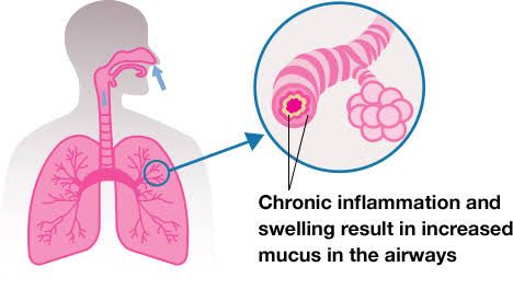
Definition of Obstructive Lung Disease
Obstructive lung disease refers to a group of respiratory conditions characterized by difficulty in exhaling air from the lungs due to airway obstruction or narrowing. This results in increased airway resistance, incomplete emptying of the lungs during expiration, and air trapping. Common examples include asthma, chronic bronchitis, bronchiectasis, and emphysema. These diseases share symptoms such as shortness of breath, wheezing, and chronic cough.
Pathogenesis, Pathological Features, and Complications
1. Asthma
- Pathogenesis: Asthma is a chronic inflammatory disorder of the airways triggered by allergens (e.g., pollen), irritants (e.g., smoke), or infections. It involves hyperresponsiveness of the bronchial smooth muscle leading to episodic bronchoconstriction. Key immune cells involved include mast cells, eosinophils, and T-helper type 2 (Th2) lymphocytes.
- Pathological Features:
- Airway inflammation with infiltration of eosinophils.
- Hypertrophy and hyperplasia of bronchial smooth muscle.
- Goblet cell hyperplasia leading to excessive mucus production.
- Thickening of the basement membrane.
- Complications:
- Status asthmaticus: A severe asthma attack that does not respond to standard treatments.
- Chronic airway remodeling resulting in irreversible airflow limitation over time.
2. Chronic Bronchitis
- Pathogenesis: Chronic bronchitis is caused by prolonged exposure to irritants like cigarette smoke or pollutants. This leads to persistent inflammation of the bronchi and excessive mucus secretion due to goblet cell hyperplasia.
- Pathological Features:
- Hyperplasia of mucus-secreting glands in the bronchi (Reid index >0.4).
- Chronic inflammation with infiltration by neutrophils and lymphocytes.
- Loss of ciliary function in the respiratory epithelium.
- Complications:
- Recurrent respiratory infections due to mucus stasis.
- Hypoxemia and hypercapnia leading to pulmonary hypertension and cor pulmonale (right-sided heart failure).
3. Bronchiectasis
- Pathogenesis: Bronchiectasis results from chronic infection or inflammation causing permanent dilation and destruction of the bronchi. It can be secondary to conditions like cystic fibrosis, tuberculosis, or recurrent pneumonia.
- Pathological Features:
- Permanent dilation of bronchi and bronchioles due to destruction of elastic tissue and smooth muscle.
- Mucopurulent secretions within dilated airways.
- Inflammatory infiltrates dominated by neutrophils in acute cases; lymphocytes may predominate chronically.
- Complications:
- Recurrent infections with organisms like Pseudomonas aeruginosa or Haemophilus influenzae.
- Hemoptysis (coughing up blood) due to erosion into adjacent blood vessels.
4. Emphysema
- Pathogenesis: Emphysema is caused by an imbalance between proteases (e.g., elastase) released by neutrophils/macrophages during inflammation and antiproteases (e.g., alpha-1 antitrypsin). Smoking is a major risk factor as it increases oxidative stress while reducing antiprotease activity. The result is destruction of alveolar walls without significant fibrosis.
- Pathological Features:
- Enlargement of airspaces distal to terminal bronchioles with loss of alveolar septa.
- Reduced surface area for gas exchange leading to hypoxemia.
- Loss of elastic recoil causing airflow obstruction during expiration.
- Complications:
- Pulmonary hypertension due to capillary bed destruction leading to right-sided heart failure (cor pulmonale).
- Pneumothorax due to rupture of bullae (large air-filled spaces).
Classification of Emphysema
Emphysema can be classified based on its morphological patterns or etiologic factors:
Morphologic Patterns
- Centriacinar Emphysema:
- Affects respiratory bronchioles predominantly in the upper lobes while sparing distal alveoli initially.
- Strongly associated with smoking.
- Panacinar Emphysema:
- Uniform enlargement from respiratory bronchioles to alveoli; affects lower lobes more commonly.
- Associated with alpha-1 antitrypsin deficiency.
- Paraseptal Emphysema:
- Involves distal acini near pleura or along interlobular septa; often seen adjacent to areas of fibrosis or scarring.
- Irregular Emphysema:
- Acini are irregularly involved; usually associated with scarring from prior infections.
Etiologic Patterns
- Smoking-related emphysema: Most common cause worldwide; primarily centriacinar pattern observed.
- Alpha-1 antitrypsin deficiency-related emphysema: Genetic condition leading predominantly to panacinar emphysema.
- Environmental/occupational exposure-related emphysema: Exposure to pollutants such as silica dust may contribute.


