Microorganisms Involved in Lower Respiratory Tract Infections (LRTIs)
Lower respiratory tract infections (LRTIs) are caused by a variety of microorganisms, including bacteria, viruses, and fungi. The most common bacterial pathogens include:
- Streptococcus pneumoniae: A leading cause of community-acquired pneumonia (CAP), bacteremia, and meningitis.
- Haemophilus influenzae: Particularly type b (Hib), which can cause pneumonia and other invasive diseases.
- Mycoplasma pneumoniae: Associated with atypical pneumonia.
- Chlamydia pneumoniae: Another cause of atypical pneumonia.
- Legionella pneumophila: Responsible for Legionnaires’ disease, a severe form of pneumonia.
- Klebsiella pneumoniae: Often associated with hospital-acquired infections and severe cases in immunocompromised individuals.
- Pseudomonas aeruginosa: Common in hospital-acquired infections, especially in patients with cystic fibrosis or chronic obstructive pulmonary disease (COPD).
- Staphylococcus aureus, including methicillin-resistant Staphylococcus aureus (MRSA): Causes severe necrotizing pneumonia.
- Moraxella catarrhalis: Frequently associated with exacerbations of COPD and secondary bacterial infections.
Viruses such as respiratory syncytial virus (RSV), influenza virus, adenovirus, and coronaviruses also play significant roles in LRTIs.
Classification of Pneumonias and the Organisms in Each Group
1. Classification by Location Acquired
Community-Acquired Pneumonia (CAP)
- CAP occurs in individuals who have not been recently hospitalized or exposed to healthcare settings.
- Common causative organisms:
- Streptococcus pneumoniae: The most common cause worldwide.
- Haemophilus influenzae: Particularly in patients with chronic obstructive pulmonary disease (COPD).
- Atypical bacteria:
- Mycoplasma pneumoniae (causes “walking pneumonia”).
- Chlamydia pneumoniae.
- Legionella pneumophila (associated with contaminated water systems).
- Viruses: Influenza virus, respiratory syncytial virus (RSV), adenovirus.
- Gram-negative bacteria: More common in at-risk populations such as elderly patients or those with comorbidities.
Hospital-Acquired Pneumonia (HAP)
- HAP develops at least 48 hours after hospital admission and is not present at the time of admission.
- Common causative organisms:
- Multidrug-resistant bacteria:
- Methicillin-resistant Staphylococcus aureus (MRSA).
- Pseudomonas aeruginosa.
- Klebsiella pneumoniae.
- Acinetobacter baumannii.
- Multidrug-resistant bacteria:
Ventilator-Associated Pneumonia (VAP)
- A subset of HAP occurring after at least 48 hours of mechanical ventilation.
- Common causative organisms:
- Similar to HAP but includes higher rates of multidrug-resistant pathogens like MRSA and Pseudomonas species.
2. Classification by Causative Organism
Typical Pneumonia
- Caused by classic bacterial pathogens that result in lobar consolidation on imaging.
- Common organisms:
- Streptococcus pneumoniae: Most frequent cause globally.
- Haemophilus influenzae.
- Klebsiella pneumoniae: Associated with alcohol use disorder and diabetes mellitus.
- Staphylococcus aureus: Often follows viral infections like influenza.
Atypical Pneumonia
- Caused by pathogens that do not typically respond to beta-lactam antibiotics and often present with diffuse interstitial infiltrates on imaging rather than lobar consolidation.
- Common organisms:
- Bacteria:
- Mycoplasma pneumoniae: Causes mild symptoms (“walking pneumonia”).
- Chlamydia pneumoniae.
- Legionella pneumophila: Associated with outbreaks from contaminated water sources.
- Coxiella burnetii: Causes Q fever, often linked to exposure to farm animals or their products.
- Chlamydia psittaci: Causes psittacosis, linked to bird exposure.
- Viruses: Influenza virus, RSV, adenovirus.
- Bacteria:
Opportunistic Pneumonia
- Occurs in immunocompromised individuals such as those with HIV/AIDS or undergoing chemotherapy.
- Common organisms:
- Fungi:
- Pneumocystis jirovecii (formerly P. carinii): A major cause in HIV/AIDS patients.
- Aspergillus species: Invasive aspergillosis in neutropenic patients.
- Cryptococcus neoformans: Seen in advanced HIV/AIDS cases.
- Bacteria:
- Nocardia species: Found in immunosuppressed individuals.
- Viruses: Cytomegalovirus (CMV), especially post-transplantation.
- Fungi:
3. Classification by Area of Lung Affected
Lobar Pneumonia
- Involves one lobe or section of a lung; typically caused by bacterial pathogens like:
- Streptococcus pneumoniae (most common).
- Less commonly, Klebsiella pneumoniae, particularly in alcoholics.
Bronchopneumonia
- Patchy involvement around bronchi and bronchioles; often caused by:
- Mixed infections involving streptococci or staphylococci.
Interstitial Pneumonia
- Involves inflammation of the lung interstitium rather than alveoli; typically caused by atypical pathogens like:
- Viruses (influenza, RSV).
- Atypical bacteria (Mycoplasma, Legionella).
4. Other Specific Types of Pneumonia
Aspiration Pneumonia
- Caused by inhalation of gastric contents or foreign material into the lungs.
- Common anaerobic bacteria involved include: –Bacteroides, Fusobacterium species.
Necrotizing Pneumonia
- Severe form characterized by liquefaction and cavitation within lung tissue; caused by pathogens such as: –Staphylococcus aureus, Klebsiella spp., Streptococcus pyogenes.
Structure of Streptococcus pneumoniae and Its Relation to Virulence, Pathogenesis, Clinical Presentation, and Vaccine Development
Structure
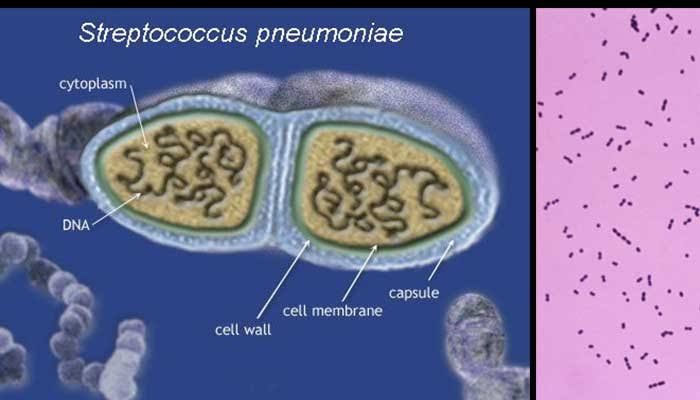
- S. pneumoniae is a Gram-positive, lancet-shaped diplococcus.
- Key structural components contributing to its virulence include:
- Polysaccharide Capsule:
- The capsule is the primary virulence factor.
- It prevents phagocytosis by inhibiting complement deposition on the bacterial surface.
- Over 100 serotypes exist; some are more virulent than others.
- Pneumolysin Toxin:
- A pore-forming toxin that damages host cells and triggers inflammation.
- Promotes bacterial dissemination from the lungs to other organs via the bloodstream.
- Surface Adhesins:
- Facilitate adherence to epithelial cells in the nasopharynx.
- Teichoic Acids and Peptidoglycan:
- Contribute to inflammation by activating immune responses.
- Polysaccharide Capsule:
Pathogenesis
S. pneumoniae colonizes the nasopharynx asymptomatically but can invade sterile sites when host defenses are compromised. Key steps include:
- Adherence to epithelial cells via adhesins.
- Evasion of immune clearance using its capsule.
- Damage to host tissues through pneumolysin-mediated cytotoxicity.
Clinical Presentation
- S. pneumoniae causes various diseases depending on where it spreads:
Vaccine Development
Vaccines target the polysaccharide capsule because it is highly immunogenic:
- Polysaccharide Vaccines (PPSV23): Protect against multiple serotypes but less effective in young children due to poor immune response to polysaccharides alone.
- Conjugate Vaccines (PCV13, PCV15, etc): Link polysaccharides to protein carriers for better immunogenicity in infants and elderly populations.
Laboratory Diagnosis and Treatment
Laboratory Diagnosis
- Specimen Collection: Samples include sputum for culture or cerebrospinal fluid for suspected meningitis cases.
- Microscopy: Gram staining reveals Gram-positive diplococci in pairs or chains.
- Culture: Grows on blood agar as alpha-hemolytic colonies under aerobic conditions; optochin sensitivity confirms identification.
- Molecular Methods: PCR detects pneumococcal DNA directly from clinical specimens for rapid diagnosis.
Treatment
- Antibiotics: First-line treatment includes beta-lactams like penicillin or amoxicillin for susceptible strains. For resistant strains or severe cases:
- Ceftriaxone or cefotaxime (third-generation cephalosporins).
- Vancomycin combined with ceftriaxone for meningitis until susceptibility results are available.
- Supportive Care: Oxygen therapy for hypoxia; fluids for dehydration; antipyretics for fever management.
- Prevention Through Vaccination: Routine use of PCV13/PCV15 vaccines has significantly reduced invasive pneumococcal disease incidence globally among children under five years old and elderly populations at risk.
Conclusion
Sufficient understanding of Streptococcus pneumoniae’s structure aids in comprehending its pathogenesis and clinical manifestations while guiding vaccine development efforts aimed at reducing global morbidity from lower respiratory tract infections.


