The Tense and Relaxed Forms of Hemoglobin
Tense (T) Form:
- The tense (T) form of hemoglobin is the deoxygenated state. In this conformation, hemoglobin has a lower affinity for oxygen.
- The T form is stabilized by salt bridges, hydrogen bonds, and hydrophobic interactions between subunits. These interactions make the structure more rigid and less accommodating to oxygen binding.
- This form is predominant in tissues where oxygen partial pressure is low, facilitating the release of oxygen to meet metabolic demands.
- Allosteric effectors such as 2,3-bisphosphoglycerate (BPG), carbon dioxide (CO2), and protons (H+) stabilize the T state further, promoting oxygen unloading.
Relaxed (R) Form:
- The relaxed (R) form of hemoglobin is the oxygenated state. In this conformation, hemoglobin has a higher affinity for oxygen.
- When one molecule of oxygen binds to a heme group in one subunit, it induces conformational changes that break some salt bridges and hydrogen bonds. This increases the affinity of other subunits for oxygen—a phenomenon known as cooperative binding.
- The R form is predominant in the lungs where oxygen partial pressure is high, allowing efficient uptake of oxygen.
Oxygen Binding Curves for Myoglobin and Hemoglobin
Myoglobin:
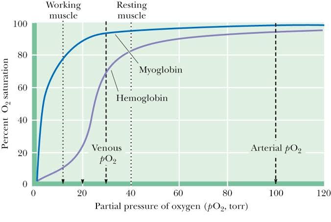
- Myoglobin exhibits a hyperbolic oxygen-binding curve because it does not exhibit cooperative binding. It consists of a single polypeptide chain with one heme group capable of binding only one molecule of oxygen.
- Myoglobin has a very high affinity for oxygen even at low partial pressures. This makes it well-suited for storing oxygen in muscle tissues but less effective at releasing it under normal physiological conditions.
Hemoglobin:
- Hemoglobin exhibits a sigmoidal (S-shaped) oxygen-binding curve due to cooperative binding among its four subunits. As each successive molecule of O2 binds to one heme group, it increases the affinity of the remaining subunits for O2.
- At low partial pressures of O2 (e.g., in tissues), hemoglobin releases O2 efficiently due to its lower affinity in the T state. At high partial pressures (e.g., in lungs), hemoglobin binds O2 efficiently due to its higher affinity in the R state.
The sigmoidal shape reflects hemoglobin’s ability to act as an efficient transporter—loading O2 in high-pressure environments like lungs and unloading it in low-pressure environments like tissues.
Equilibrium Showing Effect of O2 Binding on Tense and Relaxed Forms
The equilibrium between the Tense (T) and Relaxed (R) forms can be represented as:
(T ⇌ R)
When no or few molecules of O2 are bound:
- The equilibrium favors the T form because there are fewer conformational changes induced by bound O2.
As more molecules of O2 bind:
- Each successive binding event shifts the equilibrium toward the R form due to cooperative binding. This shift occurs because each bound O2 reduces steric hindrance and breaks stabilizing interactions that favor the T state.
This equilibrium ensures that hemoglobin transitions between states depending on environmental conditions like partial pressure of O2.
Effect of O2, BPG, CO2, and Acidity on Hemoglobin Structure and Oxygen Saturation
Oxygen:
- Oxygen acts as a positive allosteric effector by increasing hemoglobin’s affinity for additional O2 molecules through cooperative binding.
- Binding occurs preferentially when hemoglobin transitions from T to R state at high partial pressures found in alveoli.
BPG:
- BPG binds non-covalently to deoxygenated hemoglobin at a site located between β-subunits. It stabilizes the T state by reducing hemoglobin’s affinity for O2.
- This stabilization promotes greater release of O2 into tissues where it is needed most.
Carbon Dioxide:
- CO2 reacts with terminal amino groups on globin chains forming carbaminohemoglobin. This reaction stabilizes the T state by creating additional salt bridges within hemoglobin.
- Increased CO2 levels promote greater unloading of O2 into metabolically active tissues—a phenomenon known as Bohr effect.
Acidity (Protons):
- Protons bind to specific histidine residues on globin chains, stabilizing salt bridges that favor the T state.
- Lower pH levels caused by lactic acid or CO2 accumulation shift hemoglobin’s dissociation curve rightward—enhancing tissue-level delivery during acidosis or exercise.
How an Increase in BPG Helps Adaptation to High Altitude
At high altitudes:
- Atmospheric pressure decreases, leading to reduced partial pressure of oxygen (pO2).
- This results in decreased arterial O2 saturation levels since less O2 binds effectively to hemoglobin under these conditions.
Role of BPG:
- To compensate for reduced pO2, red blood cells increase production of BPG via glycolytic pathways.
- Elevated BPG levels stabilize deoxygenated hemoglobin’s T state by binding electrostatically between β-subunits.
- Stabilization lowers overall Hb-O₂-affinity while enhancing offloading capacity at tissue level despite hypoxic conditions.
This adaptation ensures sufficient delivery even when ambient pO₂-levels remain chronically low over prolonged exposure periods such mountaineering expeditions acclimatization phases alike.
Factors That Shift the Oxygen-Hemoglobin Dissociation Curve
The oxygen-hemoglobin dissociation curve can shift to the right or left depending on various physiological factors. These shifts alter hemoglobin’s affinity for oxygen, influencing oxygen transport and delivery to tissues.
Factors That Shift the Curve to the Right
A rightward shift indicates a decreased affinity of hemoglobin for oxygen, making it easier for oxygen to be released into tissues. This is beneficial in conditions where tissues require more oxygen. The following factors cause a rightward shift:
- Increased Carbon Dioxide (CO₂) Levels:
- High CO₂ levels lead to increased production of carbonic acid, lowering blood pH (Bohr effect). This stabilizes the T-state (tense state) of hemoglobin, reducing its affinity for oxygen.
- Example: During active metabolism in tissues, CO₂ levels rise, facilitating oxygen unloading.
- Decreased pH (Increased H⁺ Concentration):
- A drop in pH (acidosis) reduces hemoglobin’s affinity for oxygen by promoting protonation of certain amino acids in hemoglobin, stabilizing its deoxygenated form.
- Physiological importance: Enhances oxygen release in metabolically active tissues where lactic acid or CO₂ accumulation lowers pH.
- Increased Temperature:
- Elevated temperatures weaken the bond between hemoglobin and oxygen.
- Example: During exercise or fever, higher temperatures promote oxygen delivery to working muscles.
- Increased 2,3-Bisphosphoglycerate (2,3-BPG):
- 2,3-BPG binds preferentially to deoxyhemoglobin and reduces its affinity for oxygen.
- Physiological importance: In hypoxic conditions (e.g., high altitude), 2,3-BPG levels increase to enhance tissue oxygen delivery.
- Exercise:
- Exercise combines several factors such as increased CO₂ production, decreased pH, elevated temperature, and increased 2,3-BPG levels—all contributing to a rightward shift.
Factors That Shift the Curve to the Left
A leftward shift indicates an increased affinity of hemoglobin for oxygen, making it harder for hemoglobin to release bound oxygen into tissues. This occurs under conditions favoring enhanced loading of oxygen at the lungs:
- Decreased Carbon Dioxide Levels:
- Low CO₂ levels result in less carbonic acid formation and higher blood pH.
- Example: In hyperventilation or at rest when metabolic activity is low.
- Increased pH (Decreased H⁺ Concentration):
- Alkalosis increases hemoglobin’s affinity for oxygen by stabilizing its R-state (relaxed state).
- Physiological importance: Promotes efficient loading of oxygen at pulmonary capillaries.
- Decreased Temperature:
- Lower temperatures strengthen the bond between hemoglobin and oxygen.
- Example: In cold environments or during hypothermia.
- Decreased 2,3-BPG Levels:
- Reduced 2,3-BPG concentration increases hemoglobin’s affinity for oxygen.
- Example: Stored blood used in transfusions has lower 2,3-BPG levels due to glycolysis inhibition over time.
- Fetal Hemoglobin (HbF):
Physiological Importance of These Factors on Oxygen Transport
The shifts in the oxyhemoglobin dissociation curve are critical for maintaining efficient gas exchange under varying physiological conditions:
- A rightward shift ensures that more oxygen is released from hemoglobin into peripheral tissues during states of high metabolic demand (e.g., exercise or hypoxia). This adaptation allows tissues with low partial pressures of O₂ to receive adequate amounts despite reduced hemoglobin saturation.
- A leftward shift enhances O₂ uptake at pulmonary capillaries where partial pressure of O₂ is high but ensures less unloading at peripheral tissues unless necessary. This mechanism is crucial during rest or when metabolic demands are low.
These shifts allow dynamic regulation of O₂ delivery based on tissue needs while maintaining overall homeostasis within the body.
Shift During Exercise
During exercise, multiple physiological changes occur that collectively cause a significant rightward shift in the oxyhemoglobin dissociation curve:
- Increased CO₂ Production:
- Active muscles produce large amounts of CO₂ as a byproduct of aerobic respiration.
- The resulting increase in carbonic acid lowers blood pH via the Bohr effect.
- Decrease in Blood pH:
- Lactic acid accumulation from anaerobic metabolism further contributes to acidosis.
- Elevated Temperature:
- Muscle activity generates heat that raises local tissue temperature.
- Increased 2,3-BPG Levels:
- Prolonged exercise stimulates glycolysis in red blood cells due to hypoxia-induced signaling pathways that increase 2,3-BPG production.
These combined effects reduce hemoglobin’s affinity for O₂ significantly at peripheral tissues while maintaining effective loading at pulmonary capillaries due to high alveolar PO₂ levels during ventilation adjustments made by respiratory centers during exercise


