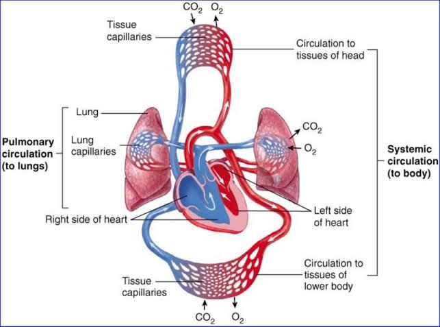
Comparison of Pulmonary and Systemic Circulations
The pulmonary and systemic circulations are two distinct components of the cardiovascular system, each serving unique roles in maintaining oxygenation and nutrient delivery throughout the body. Below is a detailed comparison:
- Primary Function:
- Pulmonary Circulation: Facilitates gas exchange by transporting deoxygenated blood from the right ventricle to the lungs, where carbon dioxide is removed, and oxygen is absorbed.
- Systemic Circulation: Delivers oxygenated blood from the left ventricle to all tissues of the body for metabolic needs and returns deoxygenated blood to the right atrium.
- Pressure Differences:
- Pulmonary Circulation: Operates at low pressure (mean pulmonary arterial pressure ~15 mmHg) due to its short distance and low resistance pathway.
- Systemic Circulation: Functions under high pressure (mean arterial pressure ~90-100 mmHg) to ensure adequate perfusion across a larger vascular network with higher resistance.
- Vessel Wall Structure:
- Pulmonary Arteries: Thin-walled with less smooth muscle, allowing for greater compliance and accommodation of cardiac output without significant increases in pressure.
- Systemic Arteries: Thick-walled with more smooth muscle and elastic fibers to withstand higher pressures.
- Blood Volume Distribution:
- Approximately 10% of total blood volume resides in the pulmonary circulation at any given time.
- The systemic circulation contains about 85% of total blood volume, reflecting its extensive vascular network.
- Response to Hypoxia:
- Pulmonary Circulation: Hypoxic pulmonary vasoconstriction occurs, redirecting blood flow away from poorly ventilated alveoli to optimize gas exchange.
- Systemic Circulation: Hypoxia typically causes vasodilation to increase oxygen delivery to tissues.
- Oxygen Content in Blood Vessels:
- In pulmonary arteries, blood is deoxygenated; in pulmonary veins, it is oxygenated.
- In systemic arteries, blood is oxygenated; in systemic veins, it is deoxygenated.
Bronchial Circulation and Physiological Shunt
Bronchial Circulation
The bronchial circulation provides oxygenated blood from the systemic circulation to support the metabolic needs of lung tissues that are not directly involved in gas exchange (e.g., conducting airways). Key features include:
- Originates from branches of the thoracic aorta or intercostal arteries.
- Supplies structures such as bronchi, connective tissue, nerves, lymph nodes, and visceral pleura.
- Venous drainage occurs via two pathways:
- One-third drains into systemic veins (e.g., azygos vein).
- Two-thirds drain into pulmonary veins, mixing with oxygenated blood returning from alveoli.
Physiological Shunt
The mixing of deoxygenated bronchial venous blood with oxygenated pulmonary venous blood creates a small “physiological shunt.” This shunt contributes slightly (~2-5%) to reducing arterial oxygen saturation because some venous admixture bypasses alveolar gas exchange.
Pressures in the Pulmonary System
The pulmonary circulation operates under significantly lower pressures compared to systemic circulation due to its shorter distance and lower vascular resistance:
- Mean Pulmonary Arterial Pressure (PAP): ~15 mmHg
- Systolic Pulmonary Arterial Pressure: ~25 mmHg
- Diastolic Pulmonary Arterial Pressure: ~8 mmHg
- Pulmonary Capillary Wedge Pressure (PCWP): ~6-12 mmHg
These low pressures are essential for preventing fluid leakage into alveoli while maintaining efficient gas exchange.
Blood Flow Through the Lungs and Its Distribution
Pathway of Blood Flow
- Deoxygenated blood exits the right ventricle via the pulmonary trunk.
- The trunk bifurcates into right and left pulmonary arteries supplying each lung.
- Blood flows through progressively smaller vessels—lobar arteries → segmental arteries → arterioles → capillaries—where gas exchange occurs at alveolar-capillary units.
- Oxygenated blood returns via venules → larger veins → four main pulmonary veins draining into the left atrium.
Distribution of Blood Flow
Blood flow within lungs is influenced by gravity, posture, ventilation-perfusion matching (V/Q ratio), and local factors like hypoxia:
- In an upright position:
- Apex (Zone 1): Lowest perfusion due to reduced hydrostatic pressure; may exhibit intermittent flow if alveolar pressure exceeds arterial pressure.
- Middle Lung (Zone 2): Moderate perfusion; flow depends on arterial-alveolar pressure gradient.
- Base (Zone 3): Highest perfusion due to increased hydrostatic pressure; continuous flow as both arterial and venous pressures exceed alveolar pressure.
This distribution ensures optimal matching between ventilation (airflow) and perfusion (blood flow), maximizing gas exchange efficiency.


