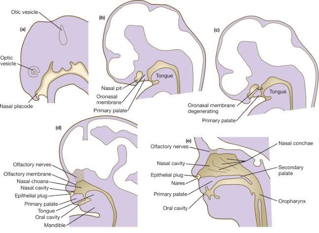
Development of the Nasal Cavity
The development of the nasal cavity begins early in embryogenesis and involves contributions from multiple germ layers. The process is as follows:
- Formation of Nasal Placodes: Around the 4th week of embryonic development, ectodermal thickenings known as nasal placodes appear on the frontonasal prominence. These placodes are critical for initiating nasal cavity formation.
- Nasal Pits Formation: By the 5th week, invagination of the nasal placodes forms depressions called nasal pits. These pits deepen to form the primitive nasal sacs, which will eventually become the nasal cavities.
- Separation from Oral Cavity: Initially, the nasal sacs are separated from the oral cavity by a thin oronasal membrane. This membrane breaks down by around the 6th week, creating a communication between the oral and nasal cavities through primitive choanae (posterior openings).
- Development of Nasal Septum and Conchae: The medial walls of the two developing nasal cavities grow downward to form a septum that divides them into right and left chambers. Lateral wall structures develop into conchae (turbinates), which increase surface area for air filtration and humidification.
- Maturation Postnatally: After birth, further growth occurs in response to environmental exposure and respiratory demands. The paranasal sinuses also expand significantly during childhood and adolescence.
Development of the Larynx
The larynx develops primarily from endodermal tissue derived from the foregut, with mesoderm contributing to its cartilaginous framework:
- Laryngotracheal Groove Formation: During Week 4, a laryngotracheal groove appears on the ventral wall of the foregut in response to signals from surrounding mesenchyme.
- Laryngotracheal Diverticulum: This groove deepens to form a laryngotracheal diverticulum, which elongates caudally while remaining connected to the pharynx at its cranial end.
- Cartilage Development: The cartilages of the larynx (thyroid, cricoid, arytenoid) arise from neural crest cells within pharyngeal arches 4 and 6 during Weeks 5–7.
- Recanalization and Vocal Fold Formation: Initially, epithelial proliferation temporarily occludes the lumen of the developing larynx by Week 10; however, recanalization occurs by Week 12–13, forming definitive vocal folds and ventricles.
- Postnatal Growth: After birth, continued growth occurs in response to vocal use and hormonal influences during puberty (especially in males).
Development of Lungs and Bronchi
The lungs and bronchi develop through distinct stages that involve branching morphogenesis:
Embryonic Stage (Weeks 4–7)
- Lung buds emerge as outgrowths from the ventral foregut endoderm around Week 4.
- These buds divide into primary bronchial buds (right and left), which push into surrounding mesenchyme covered with pleural cells.
- By Week 7, secondary bronchial buds form lobar bronchi (three on right lung; two on left lung).
Pseudoglandular Stage (Weeks 8–16)
- Extensive branching generates terminal bronchioles.
- At this stage, airways resemble exocrine glands but lack alveoli or gas exchange capability.
Canalicular Stage (Weeks 16–24)
- Terminal bronchioles give rise to respiratory bronchioles.
- Vascularization increases as capillaries approach developing airways.
- Differentiation begins between Type I alveolar cells (gas exchange) and Type II alveolar cells (surfactant production).
Saccular Stage (Week 24–Near Term)
- Respiratory bronchioles terminate in widened airspaces called saccules.
- Surfactant secretion begins around Week 26 but increases significantly closer to term.
Alveolar Stage (Late Fetal Period–Childhood)
- True alveoli begin forming late in fetal life but continue postnatally until about age eight.
- At birth, only about 15% of adult alveoli are present; postnatal growth involves both septation and expansion.
Development of Diaphragm
The diaphragm develops from multiple embryonic components that fuse together:
- Septum Transversum Contribution: The septum transversum forms early as a thickened plate of mesoderm located cranially during Weeks 3–4; it becomes part of the central tendon.
- Pleuroperitoneal Membranes: These membranes grow medially during Weeks 6–7 to close off communication between pleural and peritoneal cavities.
- Mesentery Contribution: Mesentery associated with esophagus contributes connective tissue around esophageal hiatus.
- Muscular Component from Somites: Myoblasts originating from cervical somites migrate into developing diaphragm during Weeks 5–6; these somites provide skeletal muscle fibers innervated by phrenic nerves (C3-C5).
- Closure Defects: Failure in fusion can result in congenital diaphragmatic hernias such as Bochdalek hernia.
- Postnatal Adaptation: After birth, diaphragm function matures rapidly due to its critical role in respiration.


