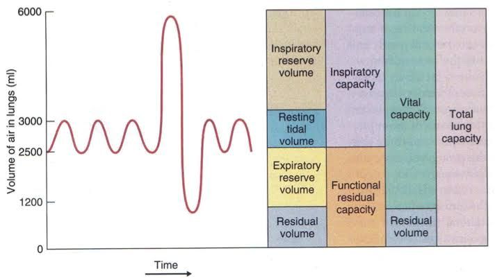
Spirometry
Spirometry is a pulmonary function test that measures lung function, specifically the volume and flow of air that can be inhaled and exhaled. It is commonly used to assess breathing patterns and diagnose respiratory conditions such as asthma, chronic obstructive pulmonary disease (COPD), pulmonary fibrosis, and cystic fibrosis. Spirometry generates spirograms, which are graphical representations of airflow (volume-time curves or flow-volume loops) during forced inhalation and exhalation. The test involves using a spirometer device to record these measurements.
Significance of the Major Volumes and Capacities Recorded During Normal Function Tests
Lung volumes and capacities provide critical insights into the mechanical properties of the lungs, chest wall, and respiratory muscles. Below are the major volumes and capacities measured in normal pulmonary function tests:
Lung Volumes
- Tidal Volume (TV):
- This is the amount of air inhaled or exhaled during a normal breath at rest.
- Normal value: Approximately 300–500 mL in adults.
- Significance: Reflects baseline respiratory activity; increases with exercise.
- Inspiratory Reserve Volume (IRV):
- The additional volume of air that can be forcibly inhaled after a normal tidal inspiration.
- Normal value: 1900–3300 mL.
- Significance: Indicates lung capacity for deep breathing.
- Expiratory Reserve Volume (ERV):
- The additional volume of air that can be forcibly exhaled after a normal tidal expiration.
- Normal value: 700–1200 mL.
- Significance: Reduced in conditions like obesity or after abdominal surgery.
- Residual Volume (RV):
- The volume of air remaining in the lungs after maximal exhalation.
- Normal value: ~1200 mL.
- Significance: Prevents lung collapse; increased in obstructive diseases like emphysema.
Lung Capacities
- Total Lung Capacity (TLC):
- The maximum volume of air the lungs can hold after maximal inspiration.
- Formula: TLC = TV + IRV + ERV + RV.
- Normal value: ~6000 mL.
- Significance: Increased in obstructive diseases; decreased in restrictive diseases.
- Vital Capacity (VC):
- The total amount of air exhaled after maximal inhalation.
- Formula: VC = TV + IRV + ERV.
- Normal value: ~4800 mL.
- Significance: Reflects inspiratory/expiratory muscle strength; reduced in restrictive disorders.
- Functional Residual Capacity (FRC):
- The volume of air remaining in the lungs at the end of a normal expiration.
- Formula: FRC = RV + ERV.
- Normal value: ~1800–2200 mL.
- Significance: Represents resting lung position; increased in COPD.
- Inspiratory Capacity (IC):
- The maximum volume of air that can be inspired following a normal expiration.
- Formula: IC = TV + IRV.
- Normal value: ~2400–3600 mL.
Algebraic Interrelations Among Pulmonary Values and Capacities
The relationships among lung volumes and capacities are algebraically defined as follows:
- TLC = TV + IRV + ERV + RV
- VC = TV + IRV + ERV
- FRC = ERV + RV
- IC = TV + IRV. These equations highlight how individual volumes combine to form larger capacities, providing insight into overall lung function.
Techniques Used to Determine Functional Residual Capacity, Residual Volume, and Total Lung Capacity
Since certain volumes like Functional Residual Capacity (FRC), Residual Volume (RV), and Total Lung Capacity (TLC) cannot be directly measured by spirometry, specialized techniques are used:
- Body Plethysmography:
- Based on Boyle’s law (P1V1 = P2V2). – A patient sits inside an airtight chamber where pressure changes during breathing allow calculation of FRC indirectly by measuring thoracic gas volume.
- Nitrogen Washout Technique:
- Involves washing out nitrogen from the lungs by having the patient breathe pure oxygen while exhaled nitrogen concentration is measured until it reaches zero.
- Helium Dilution Technique:
- Uses helium gas mixed with oxygen in a closed system to measure FRC based on helium concentration before and after equilibration with lung gases.
- Once FRC is determined: RV = FRC − ERV
TLC = VC + RV.
Closing Volume
Closing Volume refers to the lung volume at which small airways begin to close during expiration, leading to uneven ventilation within different parts of the lungs.
- It typically occurs near residual volume but increases with age or certain conditions like COPD or asthma.
- Measured using techniques such as single-breath nitrogen washout tests.
Minute Respiratory Volume
Minute Respiratory Volume is defined as the total volume of air exchanged between the lungs and atmosphere per minute under resting conditions.
- Formula: MRV = TV × Respiratory Rate
- Example: If Tidal Volume (TV) is 500 mL and Respiratory Rate is 12 breaths/minute: MRV = 500 × 12 = 6000 mL/minute or 6 L/minute.


