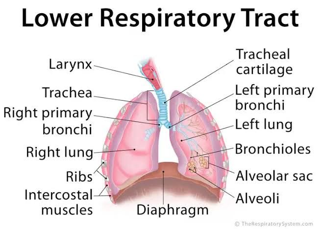
Description of the Trachea, Its Relations, and Subdivisions
The trachea, also known as the windpipe, is a tubular structure that serves as the main airway for conducting air to and from the lungs. It begins at the lower border of the cricoid cartilage (C6 vertebra) and extends down to its bifurcation at the level of the sternal angle (T4-T5 vertebral level), where it divides into the right and left primary bronchi.
Relations of the Trachea
- Anteriorly: The trachea is related to structures such as the thyroid gland (isthmus and lobes), inferior thyroid veins, sternohyoid muscles, sternothyroid muscles, and in its lower part, it is related to the arch of the aorta.
- Posteriorly: It is closely related to the esophagus throughout its length.
- Laterally: On both sides, it is flanked by the carotid sheath containing common carotid arteries, internal jugular veins, and vagus nerves. The recurrent laryngeal nerves ascend in grooves between the trachea and esophagus.
- Superiorly: It connects with the larynx via the cricotracheal ligament.
Subdivisions of the Trachea
The trachea can be divided into:
- Cervical Part: Extends from C6 to T2 vertebral levels within the neck.
- Thoracic Part: Extends from T2 to T4/T5 levels within the thorax before bifurcating into two primary bronchi.
Definition of Pleura and Pleural Cavity
Pleura
The pleura is a serous membrane that envelops each lung and lines parts of the thoracic cavity. It consists of two continuous layers:
- Visceral Pleura: Covers and adheres tightly to the surface of each lung, including its fissures.
- Parietal Pleura: Lines different parts of the thoracic cavity walls:
- Costal pleura (lines inner surfaces of ribs),
- Mediastinal pleura (covers lateral aspects of mediastinum),
- Diaphragmatic pleura (covers superior surface of diaphragm),
- Cervical pleura (extends above first rib into root of neck).
Pleural Cavity
The pleural cavity is a potential space between visceral and parietal pleurae filled with a small amount of serous fluid. This fluid reduces friction during breathing movements and creates surface tension that keeps lungs expanded against thoracic walls.
Parts and Recesses
- Costodiaphragmatic Recess: Located between costal pleura and diaphragmatic pleura; it is clinically significant as it allows lung expansion during deep inspiration.
- Costomediastinal Recess: Found between costal pleura and mediastinal pleura; more prominent on left due to cardiac notch.
Pleural Nerve Supply
The nerve supply to pleura differs based on whether it involves visceral or parietal components:
- Visceral Pleura:
- Supplied by autonomic nerves derived from pulmonary plexuses.
- Insensitive to pain but sensitive to stretch sensations.
- Parietal Pleura:
- Supplied by somatic nerves:
- Costal pleura by intercostal nerves,
- Mediastinal pleura by phrenic nerve,
- Diaphragmatic pleura by phrenic nerve centrally and lower intercostal nerves peripherally.
- Highly sensitive to pain due to somatic innervation.
- Supplied by somatic nerves:
Description of Lungs with Lobes, Fissures, Surfaces, and Comparison Between Right & Left Lungs
General Description
The lungs are paired spongy organs located in thoracic cavities on either side of mediastinum. They facilitate gas exchange between air and blood.
Each lung has:
- An apex extending above clavicle,
- A base resting on diaphragm,
- Three surfaces (costal, medial/mediastinal, diaphragmatic),
- Three borders (anterior, posterior, inferior).
Lobes & Fissures
- The right lung has three lobes separated by two fissures:
- Superior lobe,
- Middle lobe,
- Inferior lobe.
- Oblique fissure separates superior/middle lobes from inferior lobe; horizontal fissure separates superior lobe from middle lobe.
- The left lung has two lobes separated by one fissure:
- Superior lobe,
- Inferior lobe.
- Oblique fissure separates superior lobe from inferior lobe.
Additionally, left lung features a cardiac notch on anterior border for heart accommodation along with lingula—a tongue-like projection on superior lobe near cardiac notch.
Surfaces
- Costal Surface: Faces ribs; smooth due to rib impressions.
- Mediastinal Surface: Faces mediastinum; contains hilum where bronchi/vessels enter/exit lungs.
- Diaphragmatic Surface: Concave base resting on diaphragm; deeper concavity in right lung due to liver below.
List of Bronchopulmonary Segments
The lungs are divided into bronchopulmonary segments, which are the largest functional subdivisions of the lobes. Each segment is supplied by its own segmental (tertiary) bronchus and a branch of the pulmonary artery.
Right Lung
The right lung has 10 bronchopulmonary segments:
- Apical (S I)
- Posterior (S II)
- Anterior (S III)
- Lateral (S IV) – Middle lobe
- Medial (S V) – Middle lobe
- Superior (S VI) – Inferior lobe
- Medial basal (S VII) – Inferior lobe
- Anterior basal (S VIII) – Inferior lobe
- Lateral basal (S IX) – Inferior lobe
- Posterior basal (S X) – Inferior lobe
Left Lung
The left lung has 8–10 bronchopulmonary segments, as some segments may fuse:
- Apicoposterior (fusion of S I + S II)
- Anterior (S III)
- Superior lingular (S IV)
- Inferior lingular (S V)
- Superior basal (S VI)
- Anteromedial basal (fusion of S VII + S VIII in some cases)
- Lateral basal (S IX)
- Posterior basal (S X)
Innervation, Blood Supply, and Lymphatic Drainage of the Lungs
Innervation
- Parasympathetic: Derived from the vagus nerve, it stimulates secretion from bronchial glands, contraction of bronchial smooth muscle, and vasodilation of pulmonary vessels.
- Sympathetic: Derived from sympathetic trunks, it relaxes bronchial smooth muscle and causes vasoconstriction of pulmonary vessels.
- Visceral Afferent Fibers: Conduct pain impulses to the sensory ganglion of the vagus nerve.
Blood Supply
- The lungs have dual blood supply:
- Pulmonary Circulation: Pulmonary arteries deliver deoxygenated blood to the lungs for oxygenation; oxygenated blood leaves via pulmonary veins.
- Bronchial Circulation: Bronchial arteries supply oxygenated blood to lung tissues such as bronchi and visceral pleura.
- Right bronchial artery arises from the intercostobronchial trunk.
- Left bronchial arteries arise directly from the descending thoracic aorta.
Lymphatic Drainage
- Two lymphatic plexuses:
- Superficial Subpleural Plexus: Drains lung parenchyma and visceral pleura.
- Deep Plexus: Drains structures at the root of the lung.
- Both plexuses drain into tracheobronchial nodes near the tracheal bifurcation, then into right and left bronchomediastinal trunks.
Parts and Contents of the Mediastinum
The mediastinum is divided into four parts:
Superior Mediastinum
Contents include:
- Thymus
- Great vessels: Aortic arch with its branches, brachiocephalic veins, superior vena cava
- Trachea
- Esophagus
- Thoracic duct
- Vagus nerves, phrenic nerves
Anterior Mediastinum
Contents include:
- Loose connective tissue
- Fat
- Lymph nodes
Middle Mediastinum
Contents include:
- Heart enclosed in pericardium
- Ascending aorta
- Pulmonary trunk and arteries
- Pulmonary veins
- Phrenic nerves
Posterior Mediastinum
Contents include:
- Descending thoracic aorta
- Esophagus
- Thoracic duct
- Azygos vein system
Origin, Location, Course, and Branches of Internal Thoracic Artery
Origin:
The internal thoracic artery arises from the subclavian artery.
Location & Course:
It descends along both sides of the sternum within the thoracic cavity behind costal cartilages.
Branches:
- Pericardiophrenic artery
- Anterior intercostal arteries
- Musculophrenic artery
- Superior epigastric artery
Surface Markings of Trachea, Lungs, and Pleura
Trachea:
Begins at C6 vertebra level below cricoid cartilage; bifurcates at T4/T5 level at carina.
Lungs:
Apex extends above clavicle; inferior border follows rib contours—6th rib midclavicular line anteriorly; 8th rib midaxillary line laterally; 10th rib posteriorly.
Pleura:
Parietal pleura extends further than lungs—up to 12th rib posteriorly.
Typical Appearance on Chest X-Ray and CT Scan
Chest X-Ray Appearance:
- Normal lungs appear radiolucent due to air content.
- Heart shadow visible centrally with clear borders.
- Diaphragm appears dome-shaped bilaterally.
- Costophrenic angles should be sharp.
CT Scan Appearance:
- High-resolution images show detailed anatomy including bronchioles/alveoli.
- Air-filled structures appear black; soft tissues gray; bones white.
- Pathologies like tumors or fluid collections are easily visualized.


