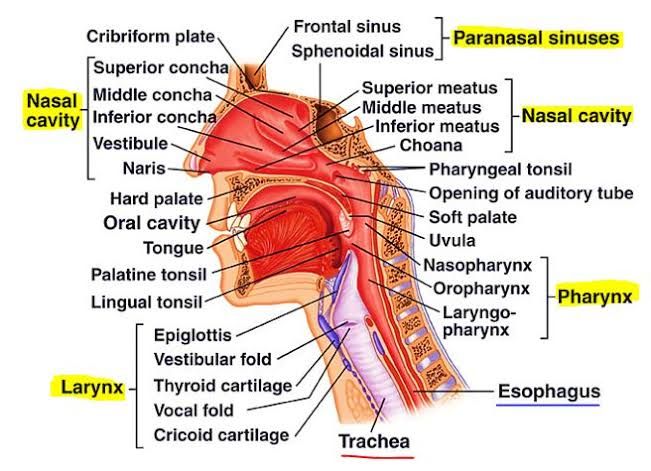
Structure of the Nasal Cavity Including Nasal Septum
The nasal cavity is the internal part of the nose, divided into two symmetrical halves by the nasal septum. It extends from the nostrils (nares) anteriorly to the choanae posteriorly, where it communicates with the nasopharynx. The nasal cavity has a roof, floor, and lateral and medial walls.
- Nasal Septum:
The nasal septum forms the medial wall of each nasal cavity and divides it into left and right halves. It consists of:- Bony components: The perpendicular plate of the ethmoid bone (superior portion) and vomer bone (posteroinferior portion).
- Cartilaginous component: The septal cartilage forms the anterior flexible part.
- Membranous part: A small area near the external nares.
- The septum is covered by mucosa that contains blood vessels and nerves for sensory innervation.
The nasal cavity is functionally divided into three regions:
- Vestibule: Located just inside the nostrils; lined with skin containing hair follicles to trap large particles.
- Respiratory region: Largest region; lined with pseudostratified ciliated columnar epithelium interspersed with goblet cells for mucus production.
- Olfactory region: Located at the apex; lined with specialized olfactory epithelium containing receptors for smell.
Structure of Lateral Wall of Nasal Cavity Including Conchae and Meatuses
The lateral wall of the nasal cavity is complex due to its bony projections called conchae (or turbinates):
- There are three conchae:
- Inferior concha (a separate bone)
- Middle concha (part of ethmoid bone)
- Superior concha (part of ethmoid bone)
These conchae create narrow air passages called meatuses:
- Inferior meatus: Located below the inferior concha.
- Middle meatus: Between inferior and middle conchae.
- Superior meatus: Between middle and superior conchae.
- Sphenoethmoidal recess: Above and behind the superior concha.
The function of these structures includes increasing surface area for humidifying, warming, and filtering inspired air while also creating turbulence to slow airflow.
Openings in Meatuses for Paranasal Sinuses and Naso-Lacrimal Duct
- Openings in each meatus allow drainage from paranasal sinuses or other structures:
- Inferior meatus: Receives drainage from the nasolacrimal duct, which carries tears from the lacrimal sac.
- Middle meatus:
- Frontal sinus drains via frontonasal duct into semilunar hiatus.
- Maxillary sinus opens into semilunar hiatus as well.
- Anterior ethmoidal cells drain here through small openings in ethmoidal bulla.
- Superior meatus: Drains posterior ethmoidal sinuses.
- Sphenoethmoidal recess: Drains sphenoid sinus.
Nasal Innervation, Blood Supply, and Relation to Epistaxis
Innervation
- Special sensory innervation for smell is provided by branches of the olfactory nerve (CN I) passing through perforations in the cribriform plate of ethmoid bone.
- General sensory innervation:
- Anterior regions are supplied by branches of ophthalmic nerve (V1, nasociliary branch).
- Posterior regions are supplied by maxillary nerve (V2, nasopalatine branch).
Blood Supply
- Rich vascular supply comes from both internal carotid artery (via anterior/posterior ethmoidal arteries) and external carotid artery (via sphenopalatine, greater palatine, superior labial, and lateral nasal arteries).
- Anastomoses occur in Kiesselbach’s plexus on the anterior septum—a common site for nosebleeds (epistaxis).
Clinical Relevance
- Epistaxis can be caused by trauma, hypertension, dry air exposure, or systemic conditions like coagulopathies. Kiesselbach’s plexus is most commonly involved in anterior epistaxis.
Structure of Nasopharynx and Associated Openings
The nasopharynx lies posterior to the nasal cavity above the soft palate. It serves as an airway connecting to both respiratory pathways.
Key Structures
- Pharyngeal tonsils (adenoids) located on its roof help trap pathogens but may obstruct airflow if enlarged.
- Openings include:
- Eustachian tube openings on lateral walls connect to middle ear for pressure equalization—clinically important as infections can spread between ear and throat via this pathway.
Structure of Cartilages and Membranes of Larynx
The larynx consists primarily of cartilage connected by membranes/ligaments:
- Unpaired cartilages:
- Thyroid cartilage (largest; forms Adam’s apple).
- Cricoid cartilage (forms a complete ring below thyroid cartilage).
- Epiglottis (elastic cartilage that prevents food entry during swallowing).
- Paired cartilages:
- Arytenoid cartilages control vocal cord tension/movement.
- Corniculate cartilages sit atop arytenoids aiding vocal fold movement.
- Cuneiform cartilages provide structural support within aryepiglottic folds.
Membranes include thyrohyoid membrane connecting thyroid cartilage to hyoid bone.
Muscles of Larynx Including Actions, Nerve Supply, Blood Supply
Intrinsic Muscles
Control vocal cords’ tension/position:
- Cricothyroid muscle tenses vocal cords (external branch of superior laryngeal nerve).
- Posterior cricoarytenoid abducts vocal cords—only muscle opening glottis fully (recurrent laryngeal nerve).
- Lateral cricoarytenoid adducts vocal cords partially closing glottis (recurrent laryngeal nerve).
Extrinsic Muscles
Elevate/depress larynx during swallowing/speech.
Nerve Supply
All intrinsic muscles except cricothyroid are innervated by recurrent laryngeal nerve; cricothyroid is supplied by external branch of superior laryngeal nerve.
Blood Supply
Largely derived from superior/inferior laryngeal arteries branching off thyroid arteries.
Structure of Vocal Cords & Mechanism of Voice Production
Structure
Vocal cords consist of true vocal folds made up of elastic fibers covered with mucosa attached between arytenoid cartilages posteriorly & thyroid cartilage anteriorly.
Mechanism
Air passes through glottis causing vibration—frequency determines pitch while amplitude affects loudness. Tension adjustments via intrinsic muscles alter pitch dynamically during speech/singing.


