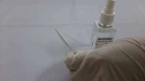
Leishman’s stain is a widely used Romanowsky stain for differentiating and identifying various types of white blood cells (leucocytes) in human blood smears. This staining technique provides clear visualization of cellular morphology, allowing for the identification of different leucocyte types based on their nuclear and cytoplasmic characteristics.
Materials Required
- Leishman Stain: A mixture of methylene blue and eosin.
- Phosphate Buffer: pH 6.8, prepared from:
- Solution A: KH2PO4 (9.1 g/L)
- Solution B: Na2HPO4 (9.5 g/L)
- Unstained Peripheral Blood Smear Slides
- Pasteur Pipette
- Kim Wipes
- Hair Dryer (optional for drying slides)
- Timer
- Cover Slips (optional for mounting)
Preparation of Phosphate Buffer
- Mix 50.8 mL of Solution A with 49.2 mL of Solution B to achieve a final volume of 100 mL at pH 6.8.
Procedure for Staining
Step 1: Preparing the Blood Smear
- Obtain a drop of fresh blood and place it on a clean glass slide.
- Use another slide to spread the blood drop evenly across the surface, creating a thin film.
- Allow the smear to air dry completely before proceeding to staining.
Step 2: Staining with Leishman Stain
- Cover the dried blood smear completely with Leishman stain using a Pasteur pipette (approximately 3 mL).
- Let the stain sit on the slide for about 2-3 minutes to allow initial dye uptake.
- Add an equal volume of phosphate buffer onto the stained slide (stain:buffer ratio = 1:1).
- Gently mix the stain and buffer by blowing over the slide with a Pasteur pipette without touching it directly.
- Leave the mixture on the slide for an additional 15-20 minutes, allowing sufficient time for staining.
Step 3: Rinsing and Drying
- After staining, rinse the slide gently under running tap water to remove excess stain.
- Wipe the back portion and edges of the slide with Kim wipes without touching the stained area.
- Optionally, dry the slide using a hair dryer set on low speed or allow it to air dry completely.
Step 4: Mounting and Viewing
- If desired, place a drop of mounting medium on top of the stained area and cover it with a clean cover slip.
- Examine under a microscope starting at low power and then switching to higher magnifications.
Interpretation of Results
- Neutrophils: Characterized by multi-lobed nuclei and granular cytoplasm that stains light pink or violet.
- Eosinophils: Identified by bi-lobed nuclei and large orange-red granules in their cytoplasm.
- Basophils: Have large dark purple granules that may obscure their lobed nuclei.
- Lymphocytes: Present as small cells with large round nuclei that take up most of their volume; cytoplasm is scanty and stains light blue.
- Monocytes: Larger cells with kidney-shaped nuclei; cytoplasm is abundant and stains gray-blue.
Conclusion
The use of Leishman’s stain allows for effective differentiation between various types of leucocytes in human blood smears due to its ability to highlight nuclear morphology and cytoplasmic details distinctly.
This comprehensive procedure ensures accurate identification and analysis of leucocyte types essential for diagnosing various hematological conditions.


