Restriction mapping, digestion, and RFLP analysis of the human HBB gene involve several systematic steps to identify genetic variations associated with this gene. The HBB gene encodes the beta-globin subunit of hemoglobin, and variations in this gene can lead to conditions such as sickle cell disease and beta-thalassemia. Below is a detailed explanation of each step involved in this process.
Step 1: Understanding the HBB Gene
The human HBB gene is located on chromosome 11 and consists of three exons separated by two introns. It plays a crucial role in encoding the beta chain of hemoglobin. Variations or mutations within this gene can significantly affect hemoglobin function and lead to various hematological disorders.
Step 2: Designing Restriction Enzymes for Digestion
To analyze the HBB gene using RFLP (Restriction Fragment Length Polymorphism), specific restriction enzymes must be selected based on their recognition sites within the HBB gene sequence. Commonly used restriction enzymes include EcoRI, HindIII, and BamHI, which recognize specific DNA sequences and cut the DNA at those sites.
- Identify Restriction Sites: The first step involves analyzing the nucleotide sequence of the HBB gene to identify potential restriction enzyme recognition sites.
- Select Enzymes: Choose enzymes that will provide informative cuts that can differentiate between alleles based on their fragment sizes.
Step 3: DNA Extraction
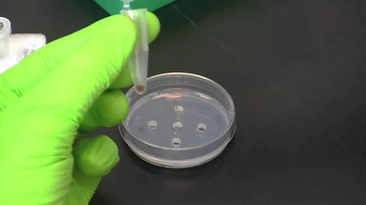
Before performing restriction digestion, genomic DNA must be extracted from human samples (e.g., blood or saliva). This process typically involves:
- Cell Lysis: Breaking open cells to release DNA.
- Purification: Removing proteins and other cellular debris using phenol-chloroform extraction or silica column methods.
- Quantification: Measuring the concentration and purity of extracted DNA using spectrophotometry.
Step 4: Restriction Digestion
Once purified genomic DNA is obtained, it undergoes digestion with selected restriction enzymes:
- Preparation of Reaction Mixture:
- Mix purified DNA with buffer solution suitable for the chosen restriction enzyme.
- Add the appropriate amount of restriction enzyme.
- Incubation:
- Incubate the reaction mixture at optimal temperature (usually around 37°C) for a specified duration (typically 1-2 hours) to allow complete digestion.
- Stopping Reaction:
- Heat inactivation may be performed by incubating at a higher temperature (e.g., 65°C) for a few minutes after digestion.
Step 5: Gel Electrophoresis
After digestion, the resulting fragments are separated by gel electrophoresis:
- Preparation of Agarose Gel:
- Prepare an agarose gel (typically 1-2% concentration) depending on fragment size expected from digestion.
- Loading Samples:
- Load digested samples into wells created in the gel along with a DNA ladder for size reference.
- Running Gel:
- Apply an electric current across the gel; smaller fragments migrate faster than larger ones, allowing separation based on size.
- Staining and Visualization:
- Stain the gel with ethidium bromide or another nucleic acid stain to visualize bands under UV light.
Step 6: Southern Blotting
To further analyze specific fragments corresponding to alleles:
- Transfer Fragments:
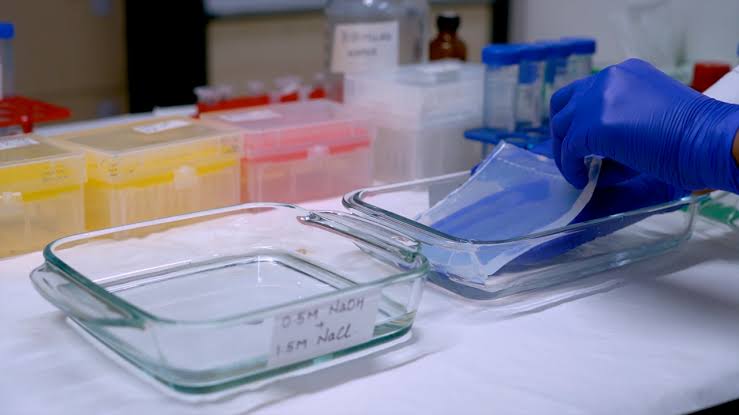
- Transfer separated DNA fragments from agarose gel onto a membrane (such as nitrocellulose) via capillary action or vacuum blotting.
- Fixing Fragments:
- Cross-link DNA to membrane using UV light or heat treatment.
Step 7: Probe Hybridization
A labeled probe complementary to a region within or near the HBB gene is used:
- Probe Preparation:
- Synthesize a labeled probe (radioactive or fluorescent) that targets specific sequences within the HBB gene region.
- Hybridization Process:
- Incubate membrane with probe solution under conditions that allow hybridization between probe and target sequences.
- Washing Steps:
- Wash away unbound probes to reduce background noise before visualization.
Step 8: Visualization and Analysis
Finally, visualize hybridized probes:
- Detection Methods:
- Use autoradiography for radioactive probes or fluorescence detection systems for fluorescent probes.
- Interpretation of Results:
- Analyze band patterns corresponding to different alleles; differences in band sizes indicate polymorphisms due to variations in restriction sites caused by mutations such as single nucleotide polymorphisms (SNPs).
- Genotype Determination:
- Determine genotypes based on observed patterns; homozygous individuals will show one band size while heterozygous individuals will show two distinct bands representing different alleles.
Conclusion
Through these systematic steps involving restriction mapping, digestion, gel electrophoresis, Southern blotting, probe hybridization, and visualization techniques, researchers can effectively analyze variations in the human HBB gene using RFLP analysis. This method not only aids in understanding genetic disorders but also has applications in genetic counseling and population genetics studies related to hemoglobinopathies like sickle cell disease and beta-thalassemia.


