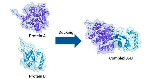
Protein–protein interactions (PPIs) refer to the specific physical contacts established between two or more protein molecules, which are crucial for various biological processes and functions within a cell. In genetic engineering, understanding PPIs is essential for manipulating genetic pathways, designing targeted therapies, and developing biotechnological applications by elucidating how proteins interact to regulate cellular functions.
(a) GST Pull-Down-Based Interaction Assay
GST Pull-Down-Based Interaction Assay is a biochemical technique used to study protein-protein interactions. In this assay, a protein of interest is fused to glutathione S-transferase (GST), which allows for the purification of the fusion protein using glutathione-coated beads. The steps involved are as follows:
- Fusion Protein Preparation: The gene encoding the target protein is cloned into a vector that expresses it as a GST fusion protein.
- Expression and Purification: The GST-fusion protein is expressed in a suitable host (often E. coli) and then purified using affinity chromatography with glutathione beads.
- Incubation with Potential Interactors: The purified GST-fusion protein is incubated with cell lysates or purified proteins that may interact with it.
- Washing: After incubation, the beads are washed to remove non-specifically bound proteins.
- Elution and Analysis: The interacting proteins are eluted from the beads, typically by adding free glutathione, and analyzed by techniques such as SDS-PAGE followed by Western blotting.
This method allows researchers to identify and characterize direct interactions between proteins.
(b) Co-Immunoprecipitation
Co-Immunoprecipitation (Co-IP) is another widely used technique for studying protein-protein interactions in biological samples. It relies on the use of antibodies to isolate a specific protein along with its interacting partners from a complex mixture, such as cell lysates. The procedure includes:
- Cell Lysis: Cells are lysed to release their contents, including proteins.
- Antibody Binding: An antibody specific to the target protein is added to the lysate, allowing it to bind to its antigen.
- Formation of Immune Complexes: Protein A/G beads (which bind antibodies) are added, forming complexes that include both the target protein and any interacting partners.
- Washing Steps: The beads are washed multiple times to remove non-specific binding proteins.
- Elution and Analysis: The immune complexes are eluted from the beads using an appropriate buffer or denaturing conditions and analyzed via SDS-PAGE and Western blotting.
Co-IP can provide insights into transient or stable interactions between proteins in their native cellular context.
(c) Yeast Two-Hybrid Assay
Yeast Two-Hybrid Assay (Y2H) is a genetic method used to detect physical interactions between two proteins within yeast cells. This assay exploits the modular nature of transcription factors, consisting of a DNA-binding domain (BD) and an activation domain (AD). Here’s how it works:
- Construction of Fusion Proteins: Two proteins of interest are each fused to either the BD or AD of a transcription factor.
- Transformation into Yeast Cells: These fusion constructs are introduced into yeast cells along with a reporter gene that can be activated by the interaction of BD and AD.
- Interaction Detection: If the two proteins interact, they bring together BD and AD, leading to activation of the reporter gene (e.g., lacZ or HIS3).
- Selection and Screening: Yeast cells that express the reporter gene can be selected on media lacking specific nutrients required for growth, indicating successful interaction.
This method allows for high-throughput screening of potential protein-protein interactions.
(d) Fluorescence Resonance Energy Transfer
Fluorescence Resonance Energy Transfer (FRET) is a powerful technique used to study molecular interactions at close range (typically 1-10 nm). FRET occurs when two fluorophores—donor and acceptor—are in proximity such that energy transfer occurs upon excitation of the donor fluorophore:
- Labeling Proteins with Fluorophores: Proteins of interest are tagged with different fluorophores; one acts as a donor (e.g., CFP) while another serves as an acceptor (e.g., YFP).
- Excitation of Donor Fluorophore: When excited by light at its specific wavelength, the donor emits fluorescence.
- Energy Transfer Mechanism: If donor and acceptor fluorophores are sufficiently close due to an interaction between their respective tagged proteins, energy from the donor can be transferred non-radiatively to the acceptor.
- Detection of FRET Signal: This transfer results in an increase in fluorescence intensity from the acceptor while decreasing intensity from the donor; this change can be quantified using specialized microscopy techniques.
FRET provides real-time information about dynamic interactions within living cells.


