Cell counting with a hemocytometer is a fundamental technique used in various biological applications, including microbiology, cell culture, and hematology. The hemocytometer allows researchers to determine the concentration of cells in a sample by providing a defined volume for counting.
Step 1: Understanding the Hemocytometer
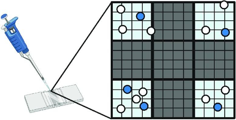
The hemocytometer is a specialized glass slide designed for counting cells. It typically features an H-shaped indentation that creates two counting chambers, each containing a grid etched onto its surface. The most commonly used type is the Improved Neubauer chamber, which has a grid subdivided into squares of known dimensions (1 mm²) and depth (0.1 mm), allowing for precise volume calculations (100 nL per square) when cells are counted.
Step 2: Preparing the Sample
To count cells accurately, it is essential to prepare the sample properly:
- Dilution: If necessary, dilute your cell suspension to achieve an optimal concentration for counting. A typical dilution factor might be 1:1 using Trypan Blue, which stains dead cells blue while live cells remain unstained.
- Mixing: Thoroughly mix the cell suspension to ensure uniform distribution before taking a sample.
Step 3: Loading the Hemocytometer
- Cleaning: Clean both the hemocytometer and coverslip with ethanol to eliminate contaminants.
- Cover Slip Placement: Place the coverslip over the counting chamber.
- Sample Loading: Using a pipette, load approximately 10 µL of your prepared cell suspension into one of the V-shaped wells of the hemocytometer. Capillary action will draw the liquid into the chamber.
Step 4: Counting Cells Under Microscope
- Microscope Setup: Place the loaded hemocytometer on the microscope stage and focus on the grid at low power (10X magnification).
- Counting Strategy:
- Count cells in designated squares (e.g., four corner squares plus one central square) to obtain an average count.
- Apply rules for counting; for example, count cells touching the top or right lines as “in” but ignore those touching bottom or left lines.
Step 5: Calculating Cell Concentration
Use this formula to calculate total cell concentration:
Total cells/mL = (Total cells counted × Dilution factor × 10,000) / Number of squares counted
For example, if you counted 325 viable cells across five squares with a dilution factor of 2:
Total cells/mL = (325 × 2 × 10,000) / 5 = 130 × 10^4
Distinguishing Different Blood Cell Types Under Microscope
When examining blood samples under a microscope using a hemocytometer:
- Staining Techniques: Use specific stains such as Wright’s stain or Giemsa stain to differentiate between various types of blood cells based on their morphology and staining characteristics.
- Identifying Cell Types:
- Red Blood Cells (RBCs): Typically appear as biconcave discs without nuclei.
- White Blood Cells (WBCs):
- Neutrophils: Multi-lobed nuclei and granular cytoplasm.
- Lymphocytes: Large nucleus with minimal cytoplasm.
- Monocytes: Kidney-shaped nucleus and abundant cytoplasm.
- Eosinophils: Bi-lobed nucleus with red granules in cytoplasm.
- Basophils: Bi-lobed nucleus with dark blue granules.
- Counting WBCs: When performing differential counts, tally each type of WBC until reaching a total count of around 100 WBCs to determine their relative proportions in the sample.
In summary, cell counting with a hemocytometer involves preparing samples accurately, loading them into a specialized chamber, systematically counting under microscopy while applying appropriate techniques for distinguishing different blood cell types based on their morphology and staining properties.


