Surface Landmarks of the Heart
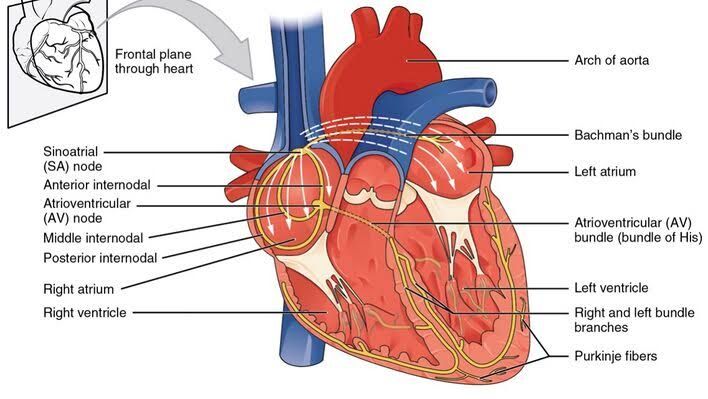
The heart is a muscular organ located in the thoracic cavity, specifically in the mediastinum, between the lungs. Its surface landmarks can be identified based on anatomical relationships and palpation points.
Location and Orientation
- Position: The heart is situated approximately behind the sternum, with its base directed upward and posteriorly, while its apex points downward and anteriorly towards the left side of the chest.
- Apex: The apex of the heart typically lies at the fifth intercostal space in adults, around 7-9 cm from the midline (the left midclavicular line).
- Base: The base of the heart is formed primarily by the left atrium and is located at approximately the level of T5-T8 vertebrae.
Borders
- Right Border: Formed mainly by the right atrium.
- Left Border: Formed by the left ventricle and a portion of the left atrium.
- Inferior Border: Primarily formed by both ventricles.
- Superior Border: Formed by both atria and great vessels.
Surface Anatomy of Great Vessels
The great vessels are major arteries and veins that enter or leave the heart, playing crucial roles in systemic and pulmonary circulation.
Major Vessels
- Aorta:
- Originates from the left ventricle.
- Ascends vertically before arching over to become the aortic arch, which then descends into the thorax as the descending aorta.
- Surface marking can be found along the right sternal border at about T2-T4 levels.
- Pulmonary Arteries:
- The right pulmonary artery branches off from the main pulmonary trunk (which arises from the right ventricle) and travels horizontally to reach each lung.
- The left pulmonary artery also arises from this trunk but runs more directly to its respective lung.
- These vessels are generally located posterior to their corresponding veins.
- Pulmonary Veins:
- Four pulmonary veins (two from each lung) return oxygenated blood to the left atrium.
- They are positioned laterally to both sides of the left atrium.
- Superior Vena Cava (SVC):
- Drains deoxygenated blood from upper body regions into the right atrium.
- It is located anteriorly at about T2-T3 levels along with its entry point into the right atrium.
- Inferior Vena Cava (IVC):
- Drains blood from lower body regions into right atrium; it enters through an opening in diaphragm at T8 level.
Surface Markings of Heart Valves and Auscultation Sites
Heart valves ensure unidirectional blood flow through chambers. Their auscultation sites can be identified on physical examination:
- Aortic Valve:
- Located at second intercostal space, right sternal border (around T2).
- Pulmonary Valve:
- Found at second intercostal space, left sternal border (around T2).
- Tricuspid Valve:
- Auscultated at fourth or fifth intercostal space along lower left sternal border (around T4).
- Mitral Valve:
- Best heard at fifth intercostal space in line with midclavicular line (around T5).
These locations correspond to where turbulent blood flow can be best detected using a stethoscope during cardiac auscultation.
Peripheral and Central Pulses for Palpation
Pulses are vital signs indicating cardiovascular health; they can be classified as peripheral or central:
Central Pulses
- Carotid Pulse:
- Located in neck beside trachea; palpated between sternocleidomastoid muscle’s anterior edge and trachea.
- Femoral Pulse:
- Found in groin area; palpated below inguinal ligament midway between pubic symphysis and anterior superior iliac spine.
Peripheral Pulses
- Radial Pulse:
- Located on wrist’s radial side; easily palpable just lateral to flexor carpi radialis tendon.
- Ulnar Pulse:
- Found on wrist’s ulnar side; less commonly palpated than radial pulse due to depth beneath tissues.
- Popliteal Pulse:
- Located behind knee joint; palpated within popliteal fossa against femur.
- Dorsalis Pedis Pulse:
- Found on dorsum of foot; palpated between first and second metatarsals near extensor hallucis longus tendon.
- Posterior Tibial Pulse:
- Located behind medial malleolus of ankle; palpated between medial malleolus and Achilles tendon.
These pulses provide essential information regarding circulation status throughout various parts of body systems.


