Ossification is the process through which bone tissue is formed. It involves the transformation of a cartilage or fibrous tissue into bone. This process is crucial for the development of the skeletal system, providing structural support and facilitating movement. There are two primary types of ossification: intramembranous ossification and endochondral ossification.
Centres of Ossification
The centres of ossification are specific locations in the body where bone formation begins. These centres can be classified into two main categories:
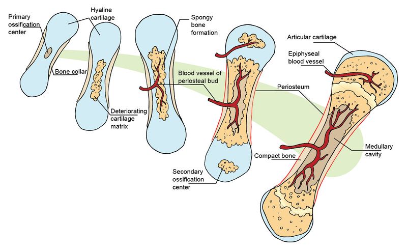
- Primary Ossification Centres: These are typically found in the diaphysis (shaft) of long bones and appear during fetal development. The primary ossification centre is where the first signs of bone formation occur, leading to the development of a bony structure from cartilage.
- Secondary Ossification Centres: These develop after birth and are usually located at the epiphyses (ends) of long bones. Secondary ossification centres contribute to the growth in length and width of bones during childhood and adolescence.
The significance of these centres lies in their role in bone growth and development, allowing for proper skeletal formation, adaptation to mechanical stress, and healing after fractures.
Intramembranous vs. Endochondral Ossification
(a) Intramembranous Ossification
Intramembranous ossification occurs directly within mesenchymal tissue (a type of connective tissue). This process is responsible for forming flat bones such as those in the skull, clavicles, and some facial bones. The steps involved include:
- Mesenchymal cells cluster together and differentiate into osteoblasts.
- Osteoblasts secrete osteoid (the organic matrix), which then mineralizes to form bone.
- As more osteoblasts surround themselves with matrix, they become osteocytes.
- The developing bone forms trabecular structures that later become compact bone.
(b) Endochondral Ossification
Endochondral ossification, on the other hand, involves a cartilage model that is gradually replaced by bone tissue. This type is responsible for forming most long bones in the body, including femur and humerus. The steps include:
- A hyaline cartilage model forms during fetal development.
- Chondrocytes within this model enlarge and die, creating cavities.
- Blood vessels invade these cavities, bringing osteoblasts that begin forming bone.
- The primary ossification centre develops in the diaphysis while secondary centres appear later in epiphyses.
- Eventually, cartilage remains only at articular surfaces and growth plates (epiphyseal plates).
Sources of Blood Supply of Long Bones
Long bones receive blood supply from several sources:
- Nutrient Artery: This major artery enters through a nutrient foramen on the diaphysis and supplies blood to the inner layers of compact bone as well as marrow.
- Metaphyseal Arteries: These arteries supply blood to the metaphysis region where active growth occurs.
- Epiphyseal Arteries: These provide blood to the epiphyses.
- Periosteal Vessels: These small vessels supply blood to the outer layer of compact bone.
This rich vascularization is essential for delivering nutrients necessary for growth, maintenance, repair processes, and overall health of bones.
Bone Markings
Bone markings refer to specific features on bones that serve various functions such as muscle attachment or articulation points with other bones:
- Projections/Processes: Such as tuberosities (large rounded projections), spines (sharp processes), or condyles (rounded ends).
- Depressions/Cavities: Such as fossae (shallow depressions) or sinuses (air-filled spaces).
- Openings: Such as foramina (holes through which nerves/vessels pass).
These markings play critical roles in biomechanics by providing leverage points for muscles or articulating surfaces for joints.
Types of Cartilages
Cartilage can be classified into three main types based on its composition and function:
- Hyaline Cartilage: Most common type; provides support with some flexibility; found in embryonic skeletons, articular surfaces, costal cartilages connecting ribs to sternum.
- Elastic Cartilage: Contains more elastic fibers than hyaline; provides strength with elasticity; found in structures like ear pinna and epiglottis.
- Fibrocartilage: Contains dense bundles of collagen fibers; provides tensile strength; found in intervertebral discs, pubic symphysis, menisci of knee joints.
These types vary significantly in their properties and functions within different anatomical contexts.


