Understanding Orthopaedic Imaging
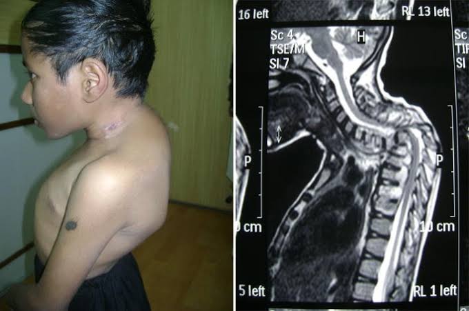
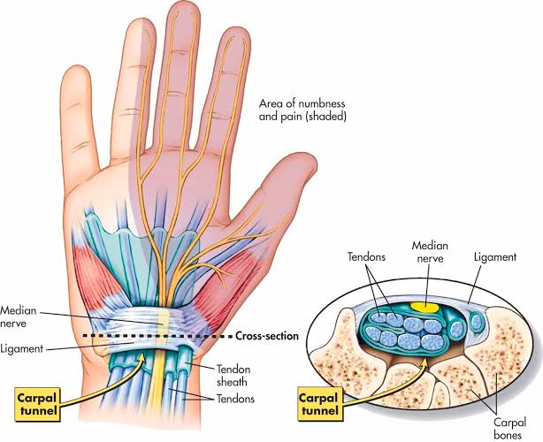
Orthopaedic imaging is crucial for diagnosing and managing musculoskeletal injuries and conditions. The primary modalities used include X-rays, MRI, CT scans, and ultrasound. Each has its specific role in the evaluation of orthopaedic issues.
Reading Orthopaedic X-Rays
Identifying Injuries
- Basic Principles: When reading an X-ray, it is essential to follow a systematic approach. This includes assessing the quality of the image (e.g., exposure, positioning) and then evaluating the anatomy.
- Common Injuries:
- Fractures: Look for discontinuity in bone cortex, alignment issues, and displacement.
- Dislocations: Identify joint spaces that are not congruent.
- Arthritis: Look for joint space narrowing, osteophyte formation, and subchondral sclerosis.
- Two Views Rule: It is standard practice to obtain at least two views (typically anteroposterior and lateral) to accurately assess fractures or dislocations since some injuries may not be visible in a single view.
- Two Joints Rule: In cases where an injury is suspected near a joint (e.g., knee or ankle), it is important to include both adjacent joints in the imaging to rule out associated injuries.
Role of Ordering Appropriate Orthopaedic X-Rays
- Indications for X-Ray:
- Suspected fractures after trauma.
- Evaluation of joint pain or swelling.
- Monitoring progression of known conditions like arthritis.
- Protocol for Ordering:
- Specify the area of concern.
- Request two views to ensure comprehensive assessment.
- Consider additional views if specific injuries are suspected (e.g., oblique views for certain fractures).
Role of MRI in Orthopaedic Practice
- Indications for MRI:
- Advantages:
- No ionizing radiation.
- Superior soft tissue contrast compared to X-rays or CT scans.
- Limitations:
- Longer scan times and higher costs.
- Not suitable for patients with certain implants or claustrophobia.
Role of CT Scans in Orthopaedic Practice
- Indications for CT Scans:
- Complex fractures (e.g., intra-articular fractures).
- Preoperative planning for surgical interventions.
- Assessment of bony lesions or tumors.
- Advantages:
- Excellent detail of bone structure.
- Rapid acquisition time compared to MRI.
- Limitations:
- Exposure to ionizing radiation.
- Less effective than MRI for soft tissue evaluation.
Role of Ultrasound in Orthopaedic Practice
- Indications for Ultrasound:
- Evaluation of soft tissue structures (tendons, ligaments).
- Guiding injections or aspirations in joints or bursae.
- Advantages:
- Real-time imaging capability allows dynamic assessment.
- No ionizing radiation; portable and cost-effective.
- Limitations:
- Operator-dependent; requires skilled personnel.
- Limited penetration through bone; not ideal for deep structures.
Conclusion
In summary, understanding how to read orthopaedic X-rays involves recognizing common injuries through systematic evaluation while adhering to protocols that ensure comprehensive imaging through multiple views when necessary. The roles of MRI, CT scans, and ultrasound complement each other by providing detailed information about soft tissues and complex bony structures that X-rays alone cannot offer.


