Division of the Tibia Bone
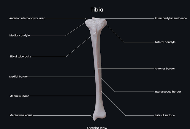
The tibia, commonly known as the shinbone, is one of the two long bones in the lower leg, the other being the fibula. The tibia can be divided into three main parts:
- Proximal End: This end of the tibia articulates with the femur at the knee joint and includes important structures such as:
- Tibial Plateau: The flat upper surface that supports the femur.
- Medial and Lateral Condyles: Rounded projections on either side of the plateau that articulate with corresponding condyles of the femur.
- Intercondylar Eminence: A bony projection between the condyles that serves as an attachment point for ligaments.
- Shaft (Diaphysis): The long, cylindrical portion of the tibia that provides structural support. It has:
- Anterior Border (Shin): A prominent ridge along the front.
- Medial Surface: Smooth and subcutaneous, allowing for easy palpation.
- Lateral Surface: More rounded and less prominent than the medial surface.
- Distal End: This end articulates with both the fibula and talus bone at the ankle joint and includes:
- Medial Malleolus: A bony prominence on the inner side of the ankle.
- Fibular Notch: A depression on its lateral aspect where it articulates with the fibula.
Surfaces and Borders of Tibia
The tibia has several distinct surfaces and borders:
- Surfaces:
- Anterior Surface: This is flat and easily palpable; it is often referred to as “the shin.”
- Medial Surface: Smooth and faces medially; it is covered by skin but can be felt easily.
- Lateral Surface: Less prominent than medial; it faces laterally towards fibula.
- Borders:
- Anterior Border (Crest): Sharp ridge running down its length; very prominent.
- Medial Border: Less defined compared to anterior border but still noticeable.
- Posterior Border (Nutrient Canal): Contains a nutrient foramen through which blood vessels enter.
Attachments of Muscles on Tibia Bone
The tibia serves as an attachment site for several muscles:
- Quadriceps Femoris Muscle Group:
- Attaches via patellar ligament at its proximal end.
- Sartorius Muscle:
- Inserts onto the medial aspect of the proximal tibia at a site known as Pes Anserinus.
- Gracilis Muscle:
- Also attaches at Pes Anserinus alongside sartorius.
- Semitendinosus Muscle:
- Another muscle attaching at Pes Anserinus.
- Tibialis Anterior Muscle:
- Originates from lateral condyle and upper two-thirds of lateral surface, inserting into medial cuneiform bone and first metatarsal.
- Soleus Muscle:
- Originates from posterior surface of head and upper quarter of shaft, contributing to plantar flexion.
- Flexor Digitorum Longus Muscle & Tibialis Posterior Muscle:
- These originate from posterior surface, aiding in toe flexion and foot inversion respectively.
Ossification of Tibia
The ossification process in bones involves two types—primary ossification centers (POCs) and secondary ossification centers (SOCs).
Primary Ossification Centers
- The primary ossification center for tibia appears during fetal development around week 7-12 gestation.
- It occurs in diaphysis where cartilage models begin to transform into bone through endochondral ossification.
Secondary Ossification Centers
- Secondary ossification centers appear after birth, primarily located at specific sites including:
- Proximal Epiphysis (around age 2-3): Contributes to growth in length until epiphyseal plates close during adolescence.
- Distal Epiphysis (around age 10-12): Also contributes to growth until closure occurs around late teens or early twenties.
These processes ensure proper growth and development of tibial structure throughout childhood into adulthood.
In summary, understanding these aspects provides insight into both functional anatomy and developmental biology related to this critical weight-bearing bone in humans.


