Parts of a Light Microscope
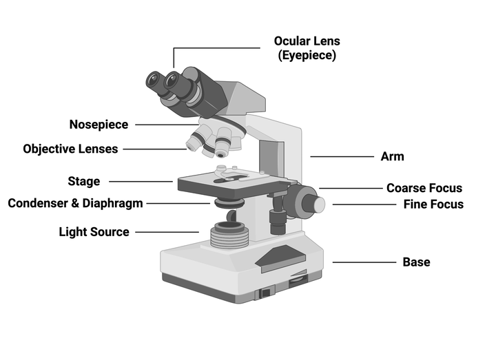
A light microscope, also known as an optical microscope, consists of several key components that work together to magnify and resolve details in specimens. The main parts include:
- Eyepiece (Ocular Lens): This is the lens at the top of the microscope through which the viewer looks. It typically has a magnification power of 10x or 15x.
- Objective Lenses: These are located on a rotating nosepiece and come in various magnifications (usually 4x, 10x, 40x, and 100x). The objective lenses gather light from the specimen and focus it to create an image.
- Stage: This is the flat platform where the slide containing the specimen is placed. It often has clips to hold the slide in place.
- Illuminator: A light source, usually a bulb or LED, that illuminates the specimen from below.
- Condenser: Located beneath the stage, this lens system focuses light onto the specimen for better illumination.
- Diaphragm: This component regulates the amount of light that reaches the specimen by adjusting an aperture.
- Focus Mechanisms: These include coarse and fine focus knobs that allow for precise adjustments to bring the specimen into clear view.
- Base and Arm: The base supports the entire microscope while the arm connects to both the base and head, providing stability.
Working of a Light Microscope
The working principle of a light microscope involves several steps:
- Illumination: The illuminator provides light that passes through a condenser lens which focuses it onto the specimen placed on the stage.
- Light Transmission: As light hits the specimen, some wavelengths are absorbed while others are transmitted or reflected depending on their properties (e.g., color, transparency).
- Magnification: The transmitted light enters through an objective lens which magnifies the image of the specimen. Depending on which objective lens is used, different levels of magnification can be achieved.
- Image Formation: The eyepiece further magnifies this image before it reaches your eye, allowing you to see details not visible to the naked eye.
- Focusing: Using coarse and fine focus knobs adjusts how close or far away these lenses are from each other and from the slide, allowing for clearer images at various depths within thicker specimens.
Magnification of Microscope
Magnification refers to how much larger an object appears under a microscope compared to its actual size. In a light microscope:
- Total Magnification = Eyepiece Magnification × Objective Lens Magnification.
For example:
- If you use a 10x eyepiece with a 40x objective lens:
Total Magnification = 10 × 40 = 400x
This means that objects appear 400 times larger than they do with just your eyes alone.
The limits of magnification in light microscopes typically range up to about 1000x due to diffraction limits imposed by visible light wavelengths; beyond this point, resolution does not improve significantly because details become indistinguishable due to overlapping wavelengths.
Different Types of Microscopes and Their Functions
There are several types of microscopes used for various applications:
- Compound Microscope: Uses multiple lenses for high magnification; ideal for viewing thin sections of specimens like tissues or cells.
- Stereo Microscope (Dissecting Microscope): Provides lower magnifications (typically up to 100x) with a three-dimensional view; useful for dissection or examining larger specimens like insects or plants.
- Phase Contrast Microscope: Enhances contrast in transparent specimens without staining; commonly used in biological research for live cell imaging.
- Fluorescence Microscope: Utilizes fluorescent dyes that emit specific colors when illuminated with particular wavelengths; widely used in cellular biology for tracking proteins or DNA within cells.
- Confocal Laser Scanning Microscope (CLSM): Uses laser beams and spatial pinholes to eliminate out-of-focus light; allows for detailed three-dimensional imaging of thick specimens such as tissues.
Types of Electron Microscopes (Transmission and Scanning)
Electron microscopes utilize electron beams instead of visible light to achieve much higher resolutions than optical microscopes:
- Transmission Electron Microscope (TEM):
- Works by transmitting electrons through ultra-thin sections of specimens.
- Capable of achieving resolutions down to atomic levels (~0.1 nm).
- Ideal for studying internal structures such as organelles within cells or materials at nanoscale dimensions.
- Scanning Electron Microscope (SEM):
- Scans a focused beam of electrons across a surface and detects secondary electrons emitted from it.
- Produces three-dimensional images with great depth-of-field but does not provide information about internal structures.
- Commonly used for examining surface morphology and texture in materials science or biological samples like pollen grains or insect exoskeletons.
In conclusion, understanding these components and functions allows researchers across various fields—from biology to materials science—to effectively utilize microscopes tailored for their specific needs while maximizing resolution and detail visibility in their observations.


