Respiratory Functions of the Lungs
- Gas Exchange: The primary function of the lungs is to facilitate the exchange of oxygen and carbon dioxide between the air and the bloodstream. This occurs in the alveoli, where oxygen from inhaled air diffuses into the blood, and carbon dioxide from the blood diffuses into the alveoli to be exhaled.
- Regulation of Blood pH: By controlling the levels of carbon dioxide in the blood through respiration, the lungs help maintain acid-base balance in the body. Increased CO2 levels can lead to a decrease in pH (acidosis), while decreased CO2 levels can increase pH (alkalosis).
- Thermoregulation: The lungs play a role in regulating body temperature through heat loss during respiration.
- Phonation: The lungs are involved in speech production by providing airflow that vibrates the vocal cords in the larynx.
Non-Respiratory Functions of the Lungs
- Defense Mechanism: The lungs trap airborne particles and pathogens, preventing them from entering systemic circulation. Particles larger than 2.5μm are generally trapped before reaching alveoli.
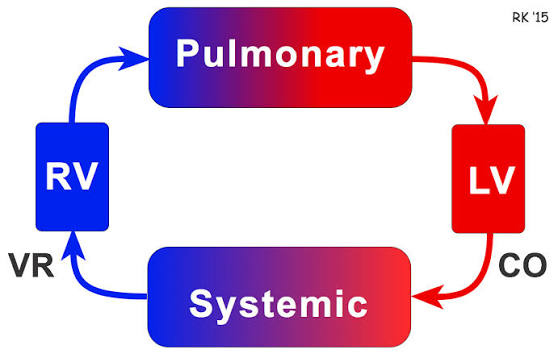
- Reservoir for Blood: The lungs contain about 10% of the circulating blood volume, acting as a reservoir that can be mobilized when needed.
- Drug Administration Route: The lungs serve as a route for administering medications, such as nebulized steroids and bronchodilators, allowing for local or systemic effects.
- Drug Elimination Route: Volatile anesthetics and other substances can be eliminated through exhalation via the lungs.
- Metabolism: The lungs participate in metabolic processes such as converting angiotensin I to angiotensin II and degrading neutrophil elastase by α1-antitrypsin.
- Modulation of Clotting Cascade: The presence of thromboplastin, heparin, and tissue plasminogen activator in lung tissue plays a role in modulating blood coagulation.
- Antimicrobial Functionality: Alveolar macrophages and other immune cells within lung tissue provide defense against infections by engulfing pathogens.
- Temperature Modulation: Heat loss occurs through respiration, contributing to overall thermoregulation.
- Filtration Function: The lungs filter out particles larger than red blood cells (~8 μm), including clots and tumor cells, thus protecting systemic circulation from potential emboli.
- Secretion of Surfactant: Although primarily related to respiratory mechanics, surfactant secretion helps reduce surface tension within alveoli, preventing collapse during exhalation.
In summary, while gas exchange is the primary function of the lungs, they also perform numerous non-respiratory functions that are crucial for overall health and homeostasis.
Nervous Control of Bronchiolar Musculature
The control of bronchiolar musculature is primarily regulated by the autonomic nervous system, which consists of both the sympathetic and parasympathetic branches. This regulation is crucial for maintaining proper airflow and respiratory function.
1. Autonomic Nervous System Overview
The autonomic nervous system operates involuntarily and is divided into two main components: the sympathetic and parasympathetic systems. Each plays a distinct role in regulating bronchiolar tone:
- Sympathetic Nervous System: This system generally promotes bronchodilation, which is the widening of the airways. It achieves this through the release of norepinephrine that binds to beta-adrenergic receptors located on the smooth muscle cells of the bronchioles. Activation of these receptors leads to relaxation of the bronchial smooth muscle, allowing for increased airflow during situations requiring heightened oxygen intake, such as exercise or stress.
- Parasympathetic Nervous System: In contrast, this system predominantly causes bronchoconstriction, which narrows the airways. The primary neurotransmitter involved is acetylcholine, released from cholinergic nerve endings. When acetylcholine binds to muscarinic receptors on bronchial smooth muscle, it induces contraction, thereby reducing airway diameter. This response can be particularly pronounced in conditions like asthma where airway hyperreactivity occurs.
2. Neurogenic Mechanisms in Asthma
In individuals with asthma, there may be abnormalities in neural control mechanisms affecting bronchiolar musculature. Research suggests that neurogenic inflammation can occur due to excitatory nonadrenergic noncholinergic (NANC) pathways that also influence airway responsiveness. These pathways can lead to increased mucus secretion and further bronchoconstriction when activated.
3. Sensory Feedback Mechanisms
The regulation of bronchiolar musculature is also influenced by sensory feedback mechanisms within the respiratory system:
- Irritant Receptors: Located in the airways, these receptors detect harmful substances or irritants and trigger reflex responses such as coughing or bronchoconstriction to protect lung tissue.
- Chemoreceptors: These are sensitive to changes in blood gases (oxygen and carbon dioxide levels) and can modulate breathing patterns accordingly.
4. Integration with Central Nervous System
The central nervous system integrates signals from various sensors throughout the body to adjust breathing patterns based on activity levels or environmental conditions. For example, during physical exertion, increased signals from peripheral chemoreceptors stimulate an increase in respiratory rate and depth through both sympathetic activation (for bronchodilation) and adjustments in parasympathetic tone.
In summary, the nervous control of bronchiolar musculature involves a complex interplay between sympathetic stimulation leading to bronchodilation and parasympathetic stimulation causing bronchoconstriction, along with sensory feedback mechanisms that help regulate these processes based on physiological needs.
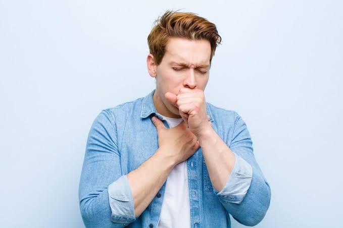
Cough Reflex Arc
The cough reflex is a protective mechanism that clears the airways of irritants through a well-defined reflex arc. The reflex arc consists of three main pathways: the sensory afferent pathway, the central pathway, and the motor efferent pathway.
- Sensory Afferent Pathway:
- The cough reflex is initiated by irritation of cough receptors located in various parts of the respiratory tract, including the trachea, bronchi, and pharynx. These receptors can be mechanoreceptors or chemoreceptors.
- There are three main types of sensory nerve fibers involved:
- Rapidly Adapting Receptors (RARs): Myelinated fibers that respond quickly to mechanical stimuli.
- Slowly Adapting Stretch Receptors (SARs): Myelinated fibers that respond more slowly and are involved in regulating lung inflation.
- C-Fibers: Non-myelinated fibers that respond to both mechanical and chemical stimuli.
- When these receptors are stimulated, they send sensory information via the vagus nerve to the medulla oblongata.
- Central Pathway:
- The sensory information travels to the nucleus tractus solitarius (NTS) in the medulla. Here, synapses occur with motor neurons that will trigger the cough reflex.
- Motor Efferent Pathway:
- The motor signals travel down various nerves to activate respiratory muscles:
- The diaphragm contracts, increasing thoracic cavity space.
- Laryngeal muscles contract to close vocal cords.
- External intercostal muscles contract to expand the chest cavity.
- Abdominal muscles may contract to assist in expelling air.
- The motor signals travel down various nerves to activate respiratory muscles:
- Phases of Cough Reflex:
- Inspiratory Phase: Vocal cords open wider; air enters lungs as external intercostal muscles and diaphragm contract.
- Compression Phase: Epiglottis and vocal cords close; pressure builds in lungs as expiration occurs against closed vocal cords.
- Expiratory Phase: Internal intercostal and abdominal muscles contract; vocal cords relax and epiglottis opens, allowing rapid expulsion of air and irritants.
Sneeze Reflex Arc
The sneeze reflex also serves as a protective mechanism for clearing irritants from the nasal passages but follows a slightly different pathway compared to coughing.
- Sensory Afferent Pathway:
- Sneezing is triggered by irritation of nasal mucosa receptors due to dust, pollen, or other irritants. These receptors include mechanoreceptors sensitive to touch or chemical stimuli.
- Sensory information is transmitted via branches of the trigeminal nerve (cranial nerve V) directly to the brainstem.
- Central Pathway:
- The sensory input reaches specific nuclei in the brainstem responsible for coordinating sneezing responses.
- Motor Efferent Pathway:
- Efferent signals travel through several cranial nerves:
- The facial nerve (cranial nerve VII) controls facial muscle contractions.
- The phrenic nerve stimulates diaphragm contraction.
- Other nerves activate abdominal muscles for forceful expiration.
- Efferent signals travel through several cranial nerves:
- Phases of Sneezing Reflex:
- Inspiratory Phase: Similar to coughing, there is an inhalation phase where air fills the lungs as vocal cords open widely.
- Compression Phase: Vocal cords close while pressure builds up in the lungs.
- Expiratory Phase: A sudden release occurs when vocal cords open rapidly, expelling air forcefully along with irritants from nasal passages.
In summary, both cough and sneeze reflexes involve complex neural pathways that initiate protective responses against irritants affecting either the lower or upper respiratory tracts respectively.


