Hyperplasia, Hypertrophy, and Atrophy of Muscle Fiber
1. Hyperplasia
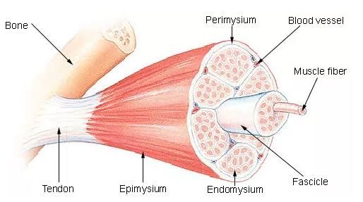
Muscle hyperplasia refers to the increase in the number of muscle fibers. This process is distinct from hypertrophy, which involves an increase in the size of existing muscle fibers. Hyperplasia is less commonly observed in human skeletal muscle compared to other species and typically occurs under specific conditions such as intense training or hormonal influences. In humans, evidence for hyperplasia is limited, but it may occur in response to certain types of resistance training or anabolic steroid use. The mechanisms behind hyperplasia involve satellite cells, which are a type of stem cell that can differentiate into new muscle fibers when stimulated by growth factors or mechanical overload.
2. Hypertrophy
Muscle hypertrophy is defined as the increase in the size of existing muscle fibers, primarily due to an increase in the synthesis of contractile proteins and other cellular components. This process occurs when the rate of protein synthesis exceeds the rate of protein degradation within the muscle cells. Hypertrophy can be induced through various forms of resistance training, particularly strength training or anaerobic exercise. There are two main types of hypertrophy: myofibrillar hypertrophy, which increases the density and size of myofibrils (the contractile units within muscle fibers), and sarcoplasmic hypertrophy, which increases the volume of sarcoplasm (the semi-fluid substance surrounding myofibrils). Factors influencing hypertrophy include mechanical tension during exercise, metabolic stress from high-repetition sets, hormonal responses (such as increased testosterone levels), and adequate nutrition.
3. Atrophy
Muscle atrophy refers to a decrease in muscle mass and strength due to a reduction in the size and number of muscle fibers. It can occur as a result of several factors including disuse (e.g., prolonged bed rest or immobilization), malnutrition, aging (sarcopenia), systemic diseases (such as cancer or chronic obstructive pulmonary disease), and neurological conditions that affect nerve supply to muscles. During atrophy, there is an imbalance where protein degradation surpasses protein synthesis, leading to a loss of contractile proteins and overall muscle volume. The extent of atrophy can vary from partial loss affecting specific muscles to complete loss affecting entire groups.
In summary:
- Hyperplasia involves an increase in fiber number.
- Hypertrophy involves an increase in fiber size.
- Atrophy involves a decrease in fiber size and strength.
Histopathological Basis of Leiomyoma
Introduction to Leiomyoma Histopathology
Uterine leiomyomas, commonly referred to as fibroids, are benign tumors that arise from the smooth muscle cells of the myometrium. The histopathological examination of leiomyomas reveals several characteristic features that differentiate them from normal uterine tissue and other neoplasms.
Microscopic Features
Histologically, leiomyomas are composed predominantly of smooth muscle cells arranged in interlacing bundles. These muscle fibers exhibit a uniform appearance without significant cellular atypia or necrosis, which is a hallmark of benign tumors. The architecture typically resembles that of normal myometrial tissue but may show variations depending on the type and location of the leiomyoma.
- Cellular Arrangement: The smooth muscle cells in leiomyomas are organized into fascicles that can be crisscrossed, mimicking the structure of normal myometrium. This arrangement contributes to the overall homogeneity observed in these tumors.
- Collagen Deposition: A notable feature in the histopathology of leiomyomas is the increased deposition of collagen within the tumor stroma. Studies have shown that interstitial collagen distribution can occupy a significant portion of the tumor area (approximately 28.53%), compared to adjacent normal tissue (around 7.43%). This excessive collagen deposition is indicative of altered extracellular matrix dynamics associated with tumor growth.
- Degenerative Changes: Leiomyomas may also exhibit degenerative changes such as hyaline degeneration, mucoid degeneration, and dystrophic calcifications. These changes can occur due to ischemia or necrosis within larger tumors or as a result of hormonal influences.
- Vascularity: Increased angiogenesis is often observed in leiomyomas, contributing to their growth and development. The presence of numerous blood vessels can be noted upon microscopic examination, which supports the metabolic demands of rapidly proliferating tumor cells.
- Immunohistochemical Markers: Immunohistochemical studies play an essential role in characterizing leiomyomas further and differentiating them from malignant counterparts like leiomyosarcomas. Common markers include estrogen receptors (ER) and progesterone receptors (PR), which are typically expressed in higher levels in leiomyomas due to their steroid hormone dependence.
Clinical Implications
The histopathological characteristics not only aid in confirming a diagnosis but also provide insights into potential clinical outcomes and treatment strategies for patients with uterine leiomyomas. Understanding these features is crucial for pathologists and clinicians alike when evaluating symptomatic patients or considering surgical interventions such as myomectomy or hysterectomy.
In summary, the histopathological basis of uterine leiomyomas encompasses distinct cellular arrangements, increased collagen deposition, degenerative changes, enhanced vascularity, and specific immunohistochemical profiles that collectively define these common benign tumors.


