Types of Smooth Muscles
Smooth muscle can be categorized into two primary types: single-unit smooth muscle and multi-unit smooth muscle. Each type has distinct structural and functional characteristics.
Single-Unit Smooth Muscle
Single-unit smooth muscle, also known as visceral smooth muscle, is the most common type of smooth muscle found in the body. Here are its key features:
- Structure: Single-unit smooth muscle cells are interconnected by gap junctions, allowing for coordinated contractions across a group of cells. This connectivity enables the entire sheet or bundle of muscle cells to contract as a single unit.
- Functionality: This type of smooth muscle exhibits myogenic activity, meaning it can generate contractions without direct neural stimulation. Instead, it responds to various stimuli such as stretch or hormonal signals. For example, the muscles in the walls of the intestines contract rhythmically to facilitate peristalsis.
- Location: Single-unit smooth muscles are predominantly found in the walls of hollow organs such as the gastrointestinal tract (stomach and intestines), bladder, uterus, and blood vessels (excluding large elastic arteries).
- Pacemaker Activity: Some cells within single-unit smooth muscles can act as pacemakers, generating rhythmic action potentials that propagate through the tissue and coordinate contractions.
Multi-Unit Smooth Muscle
Multi-unit smooth muscle is less common than single-unit smooth muscle and has different characteristics:
- Structure: In multi-unit smooth muscle, individual cells are not electrically coupled by gap junctions; instead, each cell functions independently and is innervated by autonomic nerve fibers.
- Functionality: Multi-unit smooth muscles require neural stimulation for contraction (neurogenic). The contraction is typically more precise and can be finely controlled based on specific needs rather than occurring as a synchronized wave throughout a tissue.
- Location: This type of smooth muscle is found in structures such as the iris of the eye (controlling pupil size), arrector pili muscles in hair follicles (causing hair to stand erect), and in large elastic arteries where fine control over diameter is necessary.
- Response to Stimuli: Multi-unit smooth muscles respond to neurotransmitters released from autonomic nerves, allowing for rapid adjustments in contraction based on physiological demands.
In summary, while both types of smooth muscles serve essential roles in various organ systems throughout the body, they differ significantly in their structure, functionality, and mechanisms of contraction.
Mechanism of Smooth Muscle Contraction in Comparison to Skeletal Muscle
Introduction to Muscle Types
Muscles in the human body can be categorized into three main types: skeletal, smooth, and cardiac. Each type has distinct structural and functional characteristics. Skeletal muscles are striated and under voluntary control, while smooth muscles are non-striated and operate involuntarily. Understanding the mechanisms of contraction in these muscle types reveals significant differences in their physiology.
Skeletal Muscle Contraction Mechanism
Skeletal muscle contraction is initiated by a neural signal from the motor neurons. This process involves several key steps:
- Action Potential Generation: When a motor neuron fires, it generates an action potential that travels down its axon to the neuromuscular junction.
- Release of Acetylcholine: The arrival of the action potential at the neuromuscular junction causes the release of acetylcholine (ACh) into the synaptic cleft.
- Activation of Muscle Fiber: ACh binds to receptors on the sarcolemma (muscle cell membrane), leading to depolarization and generation of an action potential in the muscle fiber.
- Calcium Release: The action potential travels along T-tubules and triggers the sarcoplasmic reticulum to release calcium ions (Ca²⁺).
- Cross-Bridge Formation: Calcium binds to troponin, causing a conformational change that moves tropomyosin away from actin binding sites. Myosin heads then attach to actin filaments, forming cross-bridges.
- Power Stroke: The myosin heads pivot, pulling actin filaments toward the center of the sarcomere, resulting in muscle contraction.
- Relaxation: When stimulation ceases, calcium is reabsorbed into the sarcoplasmic reticulum, leading to detachment of myosin from actin and relaxation of the muscle.
Smooth Muscle Contraction Mechanism
Smooth muscle contraction differs significantly from skeletal muscle contraction due to its unique structure and regulatory mechanisms:
- Initiation by Various Stimuli: Smooth muscles can be stimulated by various factors including hormones, neurotransmitters, stretch, or local chemical changes rather than solely by neural signals.
- Calcium Source: In smooth muscles, calcium ions can enter from extracellular fluid through voltage-gated calcium channels or be released from internal stores like the sarcoplasmic reticulum.
- Calmodulin Activation: Once inside the cell, Ca²⁺ binds to calmodulin (a calcium-binding protein), which activates myosin light chain kinase (MLCK).
- Myosin Phosphorylation: MLCK phosphorylates myosin light chains on myosin heads, enabling them to interact with actin filaments.
- Cross-Bridge Cycling: Similar to skeletal muscles, cross-bridge cycling occurs; however, it is regulated differently due to phosphorylation status rather than direct exposure of binding sites as seen in skeletal muscles.
- Sustained Contraction: Smooth muscle contractions can be sustained for longer periods with less energy expenditure compared to skeletal muscles due to latch state mechanisms where myosin remains attached to actin without consuming ATP continuously.
- Relaxation Mechanism: Relaxation occurs when calcium levels decrease either through reuptake into storage or extrusion out of cells; dephosphorylation of myosin light chains leads to relaxation.
Comparison Summary
In summary:
- Skeletal muscle contraction is primarily controlled by neural input and relies heavily on rapid changes in intracellular calcium levels for quick contractions.
- Smooth muscle contraction is more versatile and can be triggered by various stimuli with slower but more sustained contractions due to different regulatory mechanisms involving calmodulin and MLCK.
The differences highlight how each muscle type is adapted for its specific functions within the body—skeletal muscles for rapid movement and smooth muscles for prolonged actions such as peristalsis in digestive organs or regulating blood vessel diameter.
Physiological Anatomy of the Neuromuscular Junction in Smooth Muscle
The neuromuscular junction (NMJ) is a specialized synapse where motor neurons communicate with skeletal muscle fibers, facilitating muscle contraction. However, smooth muscle operates differently than skeletal muscle and does not have traditional neuromuscular junctions. Instead, the communication between neurons and smooth muscle fibers occurs through a series of structures and mechanisms that differ significantly from those in skeletal muscles.
1. Structure of Smooth Muscle
Smooth muscle is composed of elongated, spindle-shaped cells that are not striated like skeletal muscle fibers. Each smooth muscle cell contains a single nucleus and is surrounded by connective tissue. The arrangement of these cells allows for coordinated contractions across the entire layer of smooth muscle.
2. Innervation and Neurotransmission
Smooth muscles are innervated by autonomic motor neurons rather than somatic motor neurons as seen in skeletal muscles. The autonomic nervous system has two main divisions: the sympathetic and parasympathetic systems, which release different neurotransmitters to regulate smooth muscle activity.
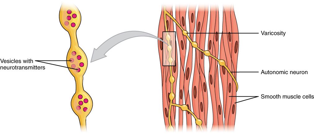
- Varicosities: Unlike the discrete NMJs found in skeletal muscles, smooth muscle fibers receive signals from varicosities—bulbous swellings along the axons of autonomic neurons. These varicosities contain neurotransmitter-filled vesicles that release their contents into the surrounding tissue rather than at a specific synaptic cleft.
- Neurotransmitters: Common neurotransmitters involved in smooth muscle contraction include acetylcholine (ACh) from parasympathetic nerves and norepinephrine from sympathetic nerves. When these neurotransmitters bind to their respective receptors on smooth muscle cells, they initiate a cascade of intracellular events leading to contraction or relaxation.
3. Receptor Types
Smooth muscle cells possess various types of receptors on their surface:
- Muscarinic Receptors: These receptors respond to acetylcholine released from parasympathetic fibers and can lead to contraction or relaxation depending on the subtype present.
- Adrenergic Receptors: These receptors respond to norepinephrine released from sympathetic fibers, influencing contraction or relaxation based on whether they are alpha or beta receptors.
4. Signal Transduction Mechanism
Upon binding of neurotransmitters to their receptors, several intracellular signaling pathways are activated:
- Calcium Ions (Ca2+): The binding of ACh or norepinephrine leads to an increase in intracellular calcium levels either through calcium influx from extracellular space or release from the sarcoplasmic reticulum.
- Calmodulin Activation: In smooth muscles, calcium binds to calmodulin instead of troponin (as in skeletal muscles). This complex activates myosin light chain kinase (MLCK), which phosphorylates myosin light chains, allowing interaction with actin filaments and resulting in contraction.
5. Relaxation Mechanisms
For relaxation to occur, calcium levels must decrease:
- Calcium Removal: Calcium is removed from the cytosol either by reuptake into the sarcoplasmic reticulum or extrusion out of the cell via pumps.
- Myosin Light Chain Phosphatase (MLCP): This enzyme dephosphorylates myosin light chains, leading to relaxation as myosin detaches from actin.
In summary, while there is no traditional neuromuscular junction in smooth muscle as seen in skeletal muscles, communication occurs through varicosities releasing neurotransmitters that interact with various receptor types on smooth muscle cells, leading to contraction or relaxation through complex signaling pathways involving calcium ions and regulatory proteins.
Types of Action Potential in Smooth Muscles
Smooth muscles exhibit two primary types of action potentials: spike potentials and action potentials with plateau. Each type plays a distinct role in the contraction and function of smooth muscle tissues.
1. Spike Potential
Spike potentials are characterized by a rapid depolarization followed by repolarization, similar to what is observed in skeletal muscle action potentials. These potentials can be elicited through various stimuli, including:
- Electrical Stimulation: Direct electrical stimulation can induce spike potentials in smooth muscle cells.
- Hormonal Influence: Certain hormones can trigger spike potentials by binding to specific receptors on the smooth muscle cell membrane.
- Neurotransmitter Release: Neurotransmitters released from nerve endings can also initiate spike potentials.
- Mechanical Stretch: The stretching of smooth muscle fibers, such as occurs in the gut during digestion, can lead to the generation of spike potentials.
The duration of spike potentials in smooth muscles is relatively short, typically lasting between 5 to 10 milliseconds. This brief action potential allows for quick contractions that are essential for various physiological processes.
2. Action Potential with Plateau
The action potential with plateau is more complex and resembles the action potential seen in cardiac muscle cells. This type of action potential is crucial for maintaining prolonged contractions within certain smooth muscles, such as those found in the uterus and ureters. Key features include:
- Prolonged Depolarization: After an initial rapid depolarization phase, there is a sustained plateau phase where the membrane remains depolarized for an extended period.
- Calcium Involvement: The plateau phase is primarily due to the influx of calcium ions (Ca²⁺) through voltage-gated calcium channels. These channels open slowly and remain open longer than sodium channels, contributing to the extended duration of contraction.
- Physiological Importance: The prolonged contraction facilitated by this type of action potential is particularly important during events like childbirth or peristalsis in the digestive tract, where sustained tension is necessary.
In summary, both spike potentials and action potentials with plateau are essential for the functionality of smooth muscles, allowing them to respond appropriately to various stimuli while facilitating necessary contractions over different time scales.
LATCH Mechanism
The LATCH mechanism refers to a physiological phenomenon observed in smooth muscle contraction. It describes a state where the muscle can maintain tension with reduced energy expenditure over time. This is particularly important during sustained contractions, such as those required for maintaining blood vessel tone or gastrointestinal motility. The mechanism is characterized by a decrease in calcium ion (Ca2+) concentration, cross-bridge phosphorylation, and ATP consumption rates while still maintaining force generation.
In smooth muscle, the contraction process involves the phosphorylation of myosin light chains, which is essential for cross-bridge attachment between actin and myosin filaments. When smooth muscle contracts initially, there are high levels of phosphorylated myosin light chains leading to strong cross-bridge cycling and rapid contraction. However, during prolonged contractions, the levels of Ca2+ and phosphorylated myosin decrease, leading to a state known as “latch.” In this state, even though the phosphorylation levels drop, the attached cross-bridges do not detach quickly due to a reduction in their detachment rate caused by dephosphorylation processes.
This latch state allows smooth muscles to maintain tension with minimal ATP consumption. The significance of this mechanism lies in its efficiency; it enables smooth muscles to sustain contractions without continuous high energy demands. This is crucial for various physiological functions such as regulating blood pressure through vascular tone and controlling peristalsis in the digestive tract.
Significance of LATCH Mechanism
The significance of the LATCH mechanism extends beyond mere energy conservation; it plays a vital role in several physiological processes:
- Energy Efficiency: By allowing smooth muscles to maintain tension with lower ATP consumption, the latch mechanism conserves energy resources within the body. This is particularly important for organs that require prolonged contractions without fatigue.
- Sustained Muscle Tone: The ability of smooth muscle to remain contracted over extended periods without continuous stimulation is essential for maintaining vascular tone and regulating blood flow throughout the circulatory system.
- Functional Adaptation: The latch mechanism allows smooth muscles to adapt their contractile properties based on physiological needs. For example, during times of increased demand (such as exercise), these muscles can quickly transition from a latch state back to active contraction when necessary.
- Pathophysiological Implications: Understanding the latch mechanism has implications for various medical conditions involving smooth muscle dysfunctions, such as hypertension or gastrointestinal disorders. Therapeutic strategies targeting this mechanism could lead to improved treatments for these conditions.
In summary, the LATCH mechanism is crucial for efficient muscle function in smooth muscle tissues, enabling sustained contractions while minimizing energy expenditure.
Nervous and Hormonal Control of Smooth Muscle Contraction
Smooth muscle contraction is primarily regulated by the autonomic nervous system (ANS) and various hormones. Understanding how these systems interact with smooth muscle is crucial for comprehending its function in the body.
1. Nervous Control
The autonomic nervous system plays a significant role in controlling smooth muscle contraction. Unlike skeletal muscles, which are controlled by well-defined neuromuscular junctions, smooth muscle fibers do not have organized motor end plates. Instead, the axons of neurons in the ANS form a series of neurotransmitter-filled bulges known as varicosities along their length. These varicosities release neurotransmitters into the synaptic cleft, allowing for communication between the nerve and the smooth muscle.
- Neurotransmitters: The primary neurotransmitters involved in smooth muscle contraction include acetylcholine (ACh) and norepinephrine (NE). ACh typically stimulates contraction in visceral smooth muscle, while NE generally causes relaxation or contraction depending on the receptor subtype present on the smooth muscle cells.
- Receptor Types: Smooth muscle cells possess various receptors that respond to different neurotransmitters. For instance, alpha-adrenergic receptors respond to norepinephrine and usually lead to contraction, while beta-adrenergic receptors can cause relaxation when activated.
- Electrical Coupling: In single-unit smooth muscles, gap junctions allow for electrical coupling between adjacent cells. This means that when one cell is stimulated by a neurotransmitter, it can spread depolarization through these junctions to neighboring cells, resulting in coordinated contractions across a larger area of tissue.
2. Hormonal Control
Hormones also play an essential role in regulating smooth muscle activity. Various hormones can influence both the contraction and relaxation of smooth muscles throughout the body.
- Hormones Involved: Some key hormones that affect smooth muscle include oxytocin, estrogen, epinephrine, and angiotensin II. For example:
- Oxytocin stimulates uterine contractions during childbirth.
- Estrogen promotes hyperplasia (increase in cell number) of uterine smooth muscle during puberty and pregnancy.
- Epinephrine, released from the adrenal glands during stress responses, can cause relaxation of certain vascular smooth muscles while inducing contraction in others depending on receptor types.
- Mechanisms of Action: Hormones exert their effects through specific receptors on smooth muscle cells. Upon binding to these receptors, they activate intracellular signaling pathways that lead to changes in calcium ion concentration within the cell or alter myosin light chain kinase activity:
- For example, when hormones bind to their respective receptors on smooth muscle cells, they may stimulate phospholipase C activity leading to increased intracellular calcium levels via release from sarcoplasmic reticulum or influx from extracellular fluid.
- Pacesetter Cells: In some regions like the gastrointestinal tract, specialized pacesetter cells can generate spontaneous action potentials that trigger contractions independent of neural input but can still be modulated by hormonal signals.
In summary, both nervous and hormonal controls are vital for regulating smooth muscle contraction. The interplay between neurotransmitters released from varicosities and hormones circulating in the bloodstream allows for fine-tuned control over involuntary movements within hollow organs and blood vessels.


