Boundaries of the Femoral Triangle
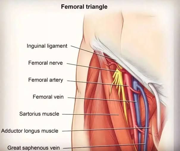
The femoral triangle is defined by three main borders, a floor, and a roof:
- Roof: The roof of the femoral triangle is formed by the fascia lata, which is a layer of connective tissue that envelops the thigh muscles.
- Floor: The floor consists of three muscles:
- Pectineus
- Iliopsoas
- Adductor longus
- Superior Border: The superior border is formed by the inguinal ligament, which extends from the anterior superior iliac spine to the pubic tubercle.
- Lateral Border: The lateral border is defined by the medial border of the sartorius muscle.
- Medial Border: The medial border is formed by the medial border of the adductor longus muscle.
It’s important to note that some sources may define the lateral border differently, but this description aligns with common anatomical references.
Contents of the Femoral Triangle
The femoral triangle contains several critical neurovascular structures arranged from lateral to medial:
- Femoral Nerve: This nerve innervates muscles in the anterior compartment of the thigh and provides sensory branches for parts of the leg and foot.
- Femoral Artery: This artery supplies blood to most of the lower limb and is a continuation of the external iliac artery as it passes under the inguinal ligament.
- Femoral Vein: Located medial to the femoral artery, this vein drains blood from the lower limb and receives drainage from the great saphenous vein within the triangle.
- Femoral Canal: This space contains deep lymph nodes and vessels and allows for distension accommodating varying levels of venous flow.
These contents are enclosed within a fascial compartment known as the femoral sheath, which helps organize these structures for efficient function and access during clinical procedures.
In summary, understanding both boundaries and contents is crucial for recognizing clinical implications such as accessing vascular structures or diagnosing conditions like femoral hernias.
Femoral Sheath and Its Compartments
The femoral sheath is a funnel-shaped fascial structure that extends from the abdomen, passing inferior to the inguinal ligament into the femoral triangle. It serves as a conduit for important neurovascular structures, specifically the femoral artery and femoral vein, allowing them to transition between the abdomen and thigh.
Structure of the Femoral Sheath
The femoral sheath is formed by an inferior extension of abdominal fascia, which includes contributions from both the transversalis fascia and iliac fascia. The sheath has a wide superior end that narrows as it descends, ultimately blending with the tunica adventitia of the femoral vessels approximately 4 cm below the inguinal ligament.
Compartments of the Femoral Sheath
The femoral sheath is divided into three distinct compartments by two vertical septae:
- Medial Compartment (Femoral Canal):
- This is the smallest compartment and contains lymphatic vessels along with a lymph node embedded in areolar tissue. It plays a role in lymphatic drainage from the lower limb.
- Intermediate Compartment:
- This compartment houses the femoral vein, which drains blood from the lower limb back to the heart. The positioning of this vein allows it to accommodate changes in venous return during movement.
- Lateral Compartment:
- This compartment contains the femoral artery, which supplies oxygenated blood to the lower limb. The artery’s location is critical for maintaining proper blood flow during activities involving hip movement.
The arrangement of these compartments within the femoral sheath facilitates efficient movement and function of both vascular structures during activities such as walking or running.
Formation of Femoral Sheath
Its formation is primarily due to the downward extension of abdominal fascia. The key components involved in its formation include:
- Abdominal Fascia: The femoral sheath originates from the transversalis fascia and psoas fascia within the abdomen. These layers of fascia extend inferiorly, creating a space that allows for the passage of important neurovascular structures.
- Inguinal Ligament: The inguinal ligament serves as a critical landmark for the femoral sheath’s location. It forms the superior boundary of this structure, with the sheath lying just below it.
- Vertical Septae: Within the femoral sheath, two vertical partitions divide it into three distinct compartments:
- The medial compartment, known as the femoral canal, contains lymphatic vessels and lymph nodes.
- The intermediate compartment houses the common femoral vein.
- The lateral compartment contains the common femoral artery.
- Termination: The inferior end of the femoral sheath blends with the tunica adventitia of both the femoral artery and vein approximately 4 cm below the inguinal ligament.
Significance of Femoral Sheath
The significance of the femoral sheath lies in its anatomical and functional roles:
- Passageway for Vessels: It provides a conduit for major blood vessels—the femoral artery and vein—to travel between the abdomen and thigh, facilitating blood circulation to and from these regions.
- Mobility During Movement: The design of the sheath allows for movement of these vessels during hip flexion and extension, which is crucial for maintaining proper blood flow during activities such as walking or running.
- Protection of Neurovascular Structures: By encasing these vessels within a fascial layer, it helps protect them from mechanical injury during movements or trauma to surrounding tissues.
- Clinical Relevance: Understanding its anatomy is essential in clinical settings, particularly in surgeries involving vascular access (such as catheterization) or in diagnosing conditions like hernias that may occur in this region.
In summary, the formation of the femoral sheath involves an intricate arrangement of abdominal fascia extending beneath the inguinal ligament into a well-defined structure that plays a vital role in vascular function and protection within the lower limb.
Formation of the Femoral Canal
The femoral canal is an anatomical structure located in the anterior thigh, specifically within the femoral sheath. It is the smallest and most medial compartment of this sheath, which also contains the femoral artery and femoral vein. The formation of the femoral canal begins with its borders:
- Medial Border: Formed by the lacunar ligament.
- Lateral Border: Defined by the femoral vein.
- Anterior Border: Created by the inguinal ligament.
- Posterior Border: Composed of the pectineal ligament, the superior ramus of the pubic bone, and the pectineus muscle.
At its superior aspect, the femoral canal opens into a space known as the femoral ring. This ring is closed off by a connective tissue layer called the femoral septum, which allows for lymphatic vessels to exit from this canal.
Contents of the Femoral Canal
The contents of the femoral canal include:
- Lymphatic Vessels: These vessels are responsible for draining lymph from deep inguinal lymph nodes.
- Deep Lymph Node: Often referred to as the lacunar node, it plays a role in immune response.
- Empty Space: This space is crucial as it allows for distension of adjacent structures, particularly accommodating increased venous return or elevated intra-abdominal pressure.
- Loose Connective Tissue: This tissue provides structural support and flexibility within the canal.
Significance of the Femoral Canal
The clinical significance of the femoral canal primarily revolves around its role in hernias, specifically femoral hernias. A hernia occurs when an internal part of the body protrudes through a weakness in surrounding tissue or muscle wall. In cases of a femoral hernia, a portion of the small intestine can push through this canal via the femoral ring.
Femoral hernias are more prevalent in women due to their wider pelvic anatomy. The rigid borders of the femoral canal can lead to compression on any herniated tissue, potentially compromising its blood supply—a condition known as strangulation. Strangulated hernias require urgent surgical intervention due to risks associated with ischemia (lack of blood flow) to affected tissues.
In summary, understanding both the formation and contents of the femoral canal is essential for recognizing its clinical implications, particularly concerning hernias that may arise in this anatomical region.
Formation of the Femoral Ring
The femoral ring is an anatomical structure located at the proximal end of the femoral canal, which is part of the femoral sheath. The formation of the femoral ring involves several key components:
- Anatomical Boundaries: The femoral ring is defined by its boundaries:
- Anteriorly, it is bounded by the inguinal ligament.
- Posteriorly, it is bordered by the pectineal ligament.
- Medially, it is limited by the crescentic base of the lacunar ligament.
- Laterally, it is adjacent to a fibrous septum that lies on the medial side of the femoral vein.
- Shape and Size: The femoral ring has an oval shape with a long diameter measuring approximately 1.25 cm. This conical opening directs superiorly and serves as a passageway for structures entering or exiting the femoral canal.
- Surrounding Structures: The femoral sheath, which contains compartments for various vessels and nerves, surrounds the femoral ring. The lateral compartment houses the femoral artery, while the intermediate compartment contains the femoral vein. The smallest compartment, known as the femoral canal, contains lymphatic vessels and lymph nodes.
- Extraperitoneal Fat: The opening of the femoral ring is filled with extraperitoneal fat that forms what is known as the femoral septum. This fat plays a role in providing cushioning and support to surrounding structures.
Significance of the Femoral Ring
The significance of the femoral ring can be understood through its clinical implications and anatomical functions:
- Pathological Relevance: One of the most critical aspects of the femoral ring is its association with hernias, particularly femoral hernias. A portion of intestinal tissue can protrude through this opening into the femoral canal, leading to incarceration or strangulation if not treated promptly.
- High Risk for Herniation: Due to its rigid boundaries—three out of four being either ligamentous or osseous—the femoral ring presents a high risk for incarceration when herniation occurs. This anatomical feature makes it less forgiving compared to other types of hernias (such as inguinal hernias), where there may be more space for movement.
- Surgical Considerations: Understanding the anatomy and significance of the femoral ring is crucial for surgeons performing procedures in this area, such as hernia repairs or vascular surgeries involving access to major blood vessels like those within the femoral sheath.
- Lymphatic Function: The presence of lymphatic vessels within the femoral canal highlights its role in immune function and fluid balance in lower limb tissues.
In summary, the formation of the femoral ring involves specific anatomical boundaries and structures that create an oval-shaped opening at the proximal end of the femoral canal, while its significance lies primarily in its association with hernias and its role in surgical anatomy.
Comparison of Anatomical Features of Femoral and Inguinal Hernias
Introduction to Hernias
Hernias occur when an internal organ or tissue bulges through a weak spot in the surrounding muscle or connective tissue. The two types of hernias being compared here are femoral hernias and inguinal hernias, both of which occur in the groin region but have distinct anatomical features.
Location
- Femoral Hernia: A femoral hernia occurs in the upper thigh, just below the groin. It involves abdominal tissue pushing through the femoral canal, which is located beneath the inguinal ligament. The femoral canal is a narrow passage that contains the femoral artery, vein, and nerve.
- Inguinal Hernia: An inguinal hernia occurs in the groin area itself. It involves part of the intestines or abdominal tissue protruding through a weakened area in the abdominal muscles, specifically through the inguinal canal. This canal is situated above the femoral canal and runs from the abdomen down to the groin.
Anatomical Structures Involved
- Femoral Hernia: The key anatomical structure involved is the femoral canal. Due to its narrowness, this type of hernia has a higher risk of incarceration (where the herniated tissue becomes trapped) and strangulation (where blood supply is cut off).
- Inguinal Hernia: The inguinal canal is composed of two parts: direct and indirect inguinal canals. Indirect inguinal hernias occur when abdominal contents pass through a congenital opening in the transversalis fascia, while direct inguinal hernias occur due to weakness in the floor of the inguinal canal.
Demographics
- Femoral Hernia: Femoral hernias are relatively uncommon and account for only 2 to 4 percent of all hernias. They are more prevalent in women, particularly older women, due to anatomical differences in pelvic structure.
- Inguinal Hernia: Inguinal hernias are much more common than femoral hernias and are significantly more prevalent in men than women. They can develop on either side but are more frequently observed on the right side.
Symptoms
- Femoral Hernia: Symptoms may include a bulge in the upper thigh near the groin, abdominal pain, nausea, vomiting, or no noticeable symptoms at all if small.
- Inguinal Hernia: Symptoms typically include a visible bulge in the groin area that may become more pronounced when standing up or coughing. Other symptoms can include burning sensations, sharp pain during physical activities like lifting or straining, and pressure or heaviness in the groin.
Conclusion
While both femoral and inguinal hernias occur in proximity to each other within the groin region, they differ significantly in their anatomical locations, structures involved, prevalence among genders, and symptomatology. Understanding these differences is crucial for diagnosis and treatment.


