Lung Volumes and Capacities
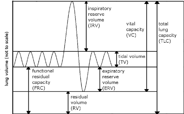
Lung volumes refer to the specific amounts of air that are inhaled or exhaled during different phases of the respiratory cycle. These measurements are crucial for assessing respiratory function and diagnosing various pulmonary conditions. The primary lung volumes include:
- Tidal Volume (TV): This is the amount of air inhaled or exhaled during a normal breath. The average adult tidal volume is approximately 300-500 mL, which constitutes about 10% of the vital capacity. It can increase significantly during exercise.
- Inspiratory Reserve Volume (IRV): This is the additional amount of air that can be forcibly inhaled after a normal tidal inspiration. The normal range for IRV in adults is between 1900-3300 mL. It reflects the lungs’ ability to take in more air when needed, such as during physical exertion.
- Expiratory Reserve Volume (ERV): This volume represents the amount of air that can be forcibly exhaled after a normal tidal expiration, typically ranging from 700-1200 mL in adults. ERV can be reduced in conditions like obesity or after abdominal surgery.
- Residual Volume (RV): This is the volume of air remaining in the lungs after maximal exhalation, averaging around 1200 mL in adults. RV cannot be measured by spirometry and is important for understanding conditions where air trapping occurs, such as obstructive lung diseases.
Lung Capacities
Lung capacities are derived from combinations of lung volumes and provide a broader view of lung function:
- Inspiratory Capacity (IC): This is the maximum volume of air that can be inhaled following a resting state and is calculated as IC = IRV + TV.
- Total Lung Capacity (TLC): This represents the total volume of air that the lungs can hold after maximum inhalation, averaging about 6 liters in adults. TLC is calculated as TLC = TV + IRV + ERV + RV and may increase in obstructive diseases like emphysema or decrease in restrictive disorders.
- Vital Capacity (VC): This measures the total amount of air exhaled after maximal inhalation, typically around 4800 mL for adults, calculated as VC = TV + IRV + ERV. VC provides insight into respiratory muscle strength and overall lung function.
- Functional Residual Capacity (FRC): This refers to the amount of air remaining in the lungs at the end of a normal expiration, calculated as FRC = RV + ERV, with normal values ranging from 1800-2200 mL. FRC helps assess lung mechanics and can indicate hyperinflation in conditions like COPD.
Clinical Significance
The measurement of these lung volumes and capacities plays an essential role in clinical practice:
- They help diagnose various pulmonary diseases by identifying restrictive or obstructive patterns.
- Changes in these values can indicate disease progression or response to treatment.
- For instance, increased residual volume may suggest obstructive lung disease due to incomplete emptying of the lungs, while decreased vital capacity may indicate restrictive disorders such as pulmonary fibrosis.
- Understanding these parameters aids clinicians in making informed decisions regarding patient management and interventions.
In summary, accurate assessment and interpretation of lung volumes and capacities are critical for evaluating respiratory health and guiding clinical care.
FEV1/FVC Ratio and Its Clinical Significance
The FEV1/FVC ratio is a critical measurement in pulmonary function testing that provides valuable insights into lung health. This ratio compares two key components of lung function: Forced Expiratory Volume in one second (FEV1) and Forced Vital Capacity (FVC). Understanding this ratio is essential for diagnosing and managing various respiratory conditions.
What is FEV1 and FVC?
- Forced Expiratory Volume in One Second (FEV1): This measures the volume of air that can be forcefully exhaled in the first second of a breath. It reflects how quickly air can be expelled from the lungs.
- Forced Vital Capacity (FVC): This measures the total amount of air that can be forcefully exhaled after taking a deep breath. It indicates the overall lung capacity.
Calculating the FEV1/FVC Ratio
The FEV1/FVC ratio is calculated by dividing the FEV1 value by the FVC value:
FEV1/FVC Ratio = FEV1 ÷ FVC
This ratio is typically expressed as a percentage. A normal FEV1/FVC ratio is generally considered to be around 70% or higher, although this can vary based on age, sex, and height.
Clinical Significance of the FEV1/FVC Ratio
The clinical significance of the FEV1/FVC ratio lies primarily in its ability to differentiate between obstructive and restrictive lung diseases:
- Obstructive Lung Diseases: Conditions such as asthma, chronic obstructive pulmonary disease (COPD), and bronchiectasis are characterized by difficulty exhaling air due to airway obstruction. In these cases, the FEV1 is reduced more than the FVC, leading to a decreased FEV1/FVC ratio (typically less than 70%). This indicates that there is an obstruction present in the airways.
- Restrictive Lung Diseases: Conditions like idiopathic pulmonary fibrosis or sarcoidosis limit lung expansion, resulting in reduced lung volumes. In restrictive diseases, both FEV1 and FVC are reduced; however, the ratio may remain normal or even increase because both values decrease proportionately. Thus, a normal or high FEV1/FVC ratio with a low FVC suggests a restrictive pattern.
- Mixed Patterns: Some patients may exhibit features of both obstructive and restrictive diseases, resulting in decreased values for both FEV1 and FVC along with a low ratio.
Monitoring Disease Progression
The FEV1/FVC ratio is not only useful for diagnosis but also plays an important role in monitoring disease progression and treatment efficacy over time. Regular spirometry tests can help healthcare providers assess whether a patient’s condition is stable, improving, or worsening.
Interpreting Results
Interpreting the results of spirometry requires consideration of demographic factors such as age, sex, height, and ethnicity since these factors influence expected values for lung function tests. Abnormal results necessitate further investigation to determine underlying causes and appropriate management strategies.
In summary, understanding the significance of the FEV1/FVC ratio aids clinicians in diagnosing respiratory conditions accurately and tailoring treatment plans effectively.
Lung Volumes and Capacities That Cannot Be Measured by Spirometer
The following lung volumes and capacities cannot be measured by spirometry:
- Residual Volume (RV) – This is the volume of air remaining in the lungs after maximal exhalation. It cannot be directly measured using a spirometer because it represents the air that is trapped in the lungs and cannot be expelled.
- Functional Residual Capacity (FRC) – This is the amount of air remaining in the lungs at the end of a normal exhalation. FRC is calculated by adding together residual volume and expiratory reserve volume, making it impossible to measure accurately with spirometry alone.
- Total Lung Capacity (TLC) – This is the maximum volume of air that the lungs can accommodate, which includes all lung volumes (TV, IRV, ERV, RV). Since TLC includes residual volume, it also cannot be measured directly by spirometry.
- Inspiratory Capacity (IC) – Although IC can be inferred from other measurements, it is not directly measurable through spirometry as it requires knowledge of both tidal volume and inspiratory reserve volume.
- Expiratory Reserve Volume (ERV) – While ERV can sometimes be estimated indirectly through spirometric techniques, its accurate measurement often requires additional methods beyond standard spirometry.
These volumes are typically assessed using alternative methods such as body plethysmography, nitrogen washout, or helium dilution techniques.
Definition of Dead Space
Dead space refers to the volume of air that is inhaled but does not participate in gas exchange within the lungs. This occurs because some of the air remains in the conducting airways or reaches alveoli that are either not perfused or poorly perfused with blood. As a result, not all the air taken in during each breath is available for the exchange of oxygen and carbon dioxide.
Types of Dead Space
- Anatomical Dead Space
- Anatomical dead space consists of the volume within the conducting airways, which includes structures from the nose and mouth down through the trachea to the terminal bronchioles. These areas conduct air to the alveoli but do not engage in gas exchange. In healthy individuals, anatomical dead space typically accounts for about 150 mL, which is roughly one-third of a normal tidal volume (450-500 mL). The volume of anatomical dead space is relatively stable and does not significantly change during various physiological states such as exercise or bronchoconstriction.
- Alveolar Dead Space
- Alveolar dead space represents those alveoli that are ventilated with fresh air but are not adequately perfused by blood flow from the pulmonary circulation. This type of dead space can increase significantly in certain lung diseases due to ventilation-perfusion mismatch, where some parts of the lung receive oxygen but do not have corresponding blood flow to facilitate gas exchange.
- Physiological Dead Space
- Physiological dead space is defined as the total dead space in an individual, which is the sum of both anatomical and alveolar dead spaces. It reflects how much inhaled air does not contribute to effective gas exchange due to either being trapped in non-exchanging areas or reaching poorly perfused alveoli.
- Mechanical (or Equipment) Dead Space
- Mechanical dead space refers to additional volumes introduced by medical equipment used during procedures such as anesthesia. This includes volumes from endotracheal tubes, masks, and other airway devices that can increase overall dead space and potentially impact ventilation efficiency.
Understanding these types of dead space is crucial for assessing respiratory function and managing conditions that affect breathing efficiency.
FEV1/FVC Ratio in Relation to Bronchial Asthma
Bronchial Asthma Overview
Bronchial asthma is a chronic inflammatory disorder of the airways characterized by variable airflow obstruction, bronchial hyperresponsiveness, and underlying inflammation. Symptoms often include wheezing, coughing, chest tightness, and shortness of breath. The pathophysiology involves complex interactions between environmental factors and genetic predisposition leading to airway inflammation and remodeling.
FEV1/FVC Ratio in Asthma Diagnosis
In individuals with asthma, the FEV1/FVC ratio can be used to determine the presence of airflow limitation. Typically, a normal FEV1/FVC ratio is around 0.75 to 0.80 in adults. In asthma patients experiencing an exacerbation or acute symptoms, this ratio may decrease below 0.70 due to increased airway resistance from bronchoconstriction and inflammation.
However, it is important to note that during periods of symptom control or remission, many asthmatics may exhibit normal FEV1/FVC ratios despite having a history of asthma. This variability underscores the need for comprehensive assessment beyond just this ratio.
Variability in FEV1/FVC Ratios
Asthma is characterized by its episodic nature; therefore, repeated measurements are often necessary for accurate diagnosis and management. The FEV1 can fluctuate significantly based on various factors such as allergen exposure, respiratory infections, physical activity, and medication adherence. Consequently, clinicians often rely on peak flow monitoring alongside spirometry results to capture these fluctuations effectively.
Implications for Treatment
The interpretation of the FEV1/FVC ratio has implications for treatment strategies in asthma management. A reduced ratio indicates more severe obstruction which may necessitate higher doses of bronchodilators or corticosteroids for effective control. Conversely, maintaining a normal FEV1/FVC ratio suggests adequate control over asthma symptoms with current treatment regimens.
Conclusion
In summary, the FEV1/FVC ratio serves as an essential tool in diagnosing and managing bronchial asthma but must be interpreted within the broader context of clinical symptoms and other diagnostic tests. Understanding its role helps healthcare providers tailor treatment plans effectively for individuals with this chronic condition.
FEV1/FVC Ratio in Relation to Chronic Obstructive Pulmonary Disease and Restrictive Lung Diseases
The FEV1/FVC ratio is calculated by dividing the FEV1 value by the FVC value. This ratio helps determine whether a patient has an obstructive or restrictive lung disease:
- Obstructive Lung Diseases: In conditions such as Chronic Obstructive Pulmonary Disease (COPD) and asthma, there is an obstruction in airflow due to inflammation, mucus production, or airway constriction. Patients with these conditions typically exhibit a reduced FEV1 with a relatively preserved or less affected FVC. As a result, the FEV1/FVC ratio falls below the normal threshold of 70%. This indicates that while patients can still take in air (as reflected by their FVC), they struggle to expel it quickly due to airway obstruction.
- Restrictive Lung Diseases: In contrast, restrictive lung diseases such as pulmonary fibrosis or sarcoidosis involve a reduction in lung volume due to stiffness in the lungs or chest wall. In these cases, both FEV1 and FVC are reduced proportionally; thus, the FEV1/FVC ratio may remain normal or even be elevated (>70%). This indicates that although patients have difficulty filling their lungs completely (reduced total lung capacity), they can still expel air effectively relative to their total lung capacity.
Clinical Implications of the FEV1/FVC Ratio
The differentiation between obstructive and restrictive patterns using the FEV1/FVC ratio has significant clinical implications:
- Diagnosis: A low ratio (<70%) suggests an obstructive pattern typical of COPD or asthma. Conversely, a normal or high ratio suggests a restrictive pattern.
- Severity Assessment: For COPD specifically, further classification into GOLD stages relies on both absolute values of FEV1 and the percentage predicted based on demographic factors like age, height, gender, and ethnicity. The new STAR classification system also utilizes this ratio for better discrimination among severity stages.
- Monitoring Disease Progression: Regular spirometry tests help monitor changes in lung function over time for both obstructive and restrictive diseases. A decline in either parameter can indicate worsening disease status.
Conclusion
In summary, the FEV1/FVC ratio serves as a vital tool for distinguishing between chronic obstructive pulmonary disease and restrictive lung diseases. Understanding this relationship aids healthcare providers in diagnosing conditions accurately and tailoring appropriate management strategies for patients.
FEV1/FVC Ratio in Relation to Pulmonary Embolism
Pulmonary Embolism Overview
Pulmonary embolism occurs when a blood clot travels to the lungs, blocking a pulmonary artery. This blockage can lead to various respiratory symptoms, including shortness of breath, chest pain, and hypoxemia. The physiological impact of PE on lung function can vary based on the size and location of the embolus, as well as the patient’s overall health.
Impact on FEV1/FVC Ratio
In patients with pulmonary embolism:
- Acute Phase Effects: During an acute PE event, patients may experience decreased lung perfusion and ventilation-perfusion mismatch. This can lead to hypoxemia and increased work of breathing. However, these changes do not typically manifest as a classic obstructive pattern characterized by a reduced FEV1/FVC ratio. Instead, patients may maintain a normal or slightly altered FEV1/FVC ratio because PE does not primarily cause airway obstruction but rather affects gas exchange efficiency.
- Chronic Effects: In cases where PE leads to chronic thromboembolic pulmonary hypertension (CTEPH), there may be more significant long-term effects on lung function. CTEPH can result in restrictive patterns due to vascular remodeling and decreased lung compliance over time. In such cases, patients might exhibit a reduced FVC while maintaining relatively preserved FEV1 levels, leading to an increased FEV1/FVC ratio (>70%), which indicates a restrictive defect rather than an obstructive one.
- Differentiation from Other Conditions: It is essential to differentiate PE from other conditions that might present with similar symptoms but have distinct impacts on lung function tests. For instance, asthma or chronic obstructive pulmonary disease (COPD) would show an obstructive pattern with a decreased FEV1/FVC ratio (<70%).
- Clinical Implications: Clinicians should interpret PFT results cautiously in patients suspected of having PE. While PFTs are valuable for assessing overall lung function and guiding management strategies for chronic respiratory diseases, they are less definitive for diagnosing acute PE directly.
In summary, while the FEV1/FVC ratio provides essential information about lung mechanics, its interpretation in relation to pulmonary embolism must consider the acute versus chronic phases of the disease and how it affects airflow dynamics differently compared to traditional obstructive or restrictive lung diseases.


