Cutaneous Nerve Supply and Dermatomes of the Thorax
The thorax, or chest region, is supplied by a complex network of cutaneous nerves that provide sensory innervation to the skin. The primary sources of this nerve supply are the thoracic spinal nerves, which emerge from the spinal cord and branch out to form various peripheral nerves. Each thoracic spinal nerve corresponds to specific dermatomes that cover distinct areas of the thoracic skin.
Thoracic Spinal Nerves and Their Role
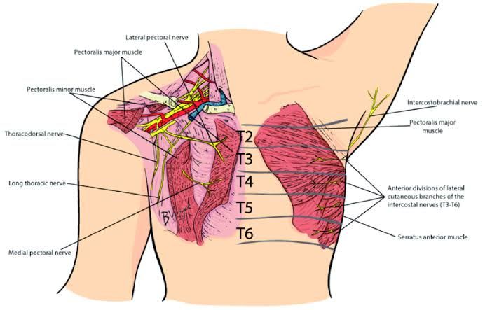
There are 12 pairs of thoracic spinal nerves (T1-T12), each contributing to the cutaneous innervation of the thorax. These nerves arise from the spinal cord and exit through intervertebral foramina. The first thoracic nerve (T1) primarily supplies the upper part of the chest, while subsequent nerves progressively innervate lower regions.
Dermatome Distribution in the Thorax
- T1 Dermatome: This dermatome covers a small area in the axilla (armpit) and medial aspect of the arm.
- T2 Dermatome: It extends over the upper part of the chest wall and includes parts of the axilla.
- T3 Dermatome: This dermatome covers a band across the upper thorax at approximately nipple level.
- T4 Dermatome: It corresponds to an area around the nipples.
- T5 Dermatome: This dermatome extends over a region just below T4, covering parts of the lower chest.
- T6 Dermatome: It continues downward, covering areas below T5 and above T7.
- T7-T9 Dermatomes: These dermatomes cover more inferior portions of the thorax, with T8 being around the xiphoid process level.
- T10 Dermatome: This dermatome corresponds to an area around the umbilicus (navel).
- T11-T12 Dermatomes: These cover lower abdominal areas and may extend into parts of the groin.
Each dermatome overlaps slightly with adjacent dermatomes, allowing for some redundancy in sensory innervation. This overlap is crucial for maintaining sensation even if one nerve root is compromised.
Clinical Significance
Understanding dermatomes is essential for diagnosing conditions affecting spinal nerves or related structures in clinical practice. For instance, if a patient presents with loss of sensation or pain in a specific area corresponding to a particular dermatome, it can indicate issues such as radiculopathy or other neurological disorders affecting that specific spinal nerve root.
In summary, cutaneous nerve supply in the thorax is primarily derived from thoracic spinal nerves T1 through T12, each responsible for distinct dermatomal areas on the chest wall.
Anatomical Justification of the Manifestations of Herpes Zoster Infection on Thoracic Wall
Herpes zoster, commonly known as shingles, is caused by the reactivation of the Varicella-zoster virus (VZV), which remains dormant in the sensory ganglia after an individual has recovered from chickenpox. The anatomical justification for the manifestations of herpes zoster on the thoracic wall can be understood through several key points:
1. Sensory Nerve Distribution: The thoracic wall is innervated by spinal nerves that emerge from the thoracic segments of the spinal cord (T1-T12). Each spinal nerve gives rise to a dorsal root that carries sensory information from specific dermatomes. When VZV reactivates, it travels down these sensory nerves and manifests along their corresponding dermatomes. The most common presentation of herpes zoster occurs in one or two adjacent dermatomes, particularly affecting the thoracic region.
2. Dermatomal Pattern: The rash associated with herpes zoster typically follows a dermatomal distribution, which means it appears in a band-like pattern along the skin supplied by a single spinal nerve root. In cases where herpes zoster affects the thoracic wall, it usually presents unilaterally and does not cross the midline due to the segmental nature of nerve innervation. This characteristic pattern is crucial for diagnosis and reflects how VZV reactivates within specific sensory ganglia.
3. Pathophysiology of Viral Reactivation: The reactivation of VZV can occur due to various factors such as stress, immunosuppression, or aging. Once reactivated, VZV travels along axons to peripheral tissues where it causes inflammation and vesicular lesions. The inflammatory response leads to pain and discomfort in addition to the characteristic rash. The thoracic wall’s anatomy allows for localized symptoms because each dermatome corresponds to a specific area of skin innervated by its respective spinal nerve.
4. Clinical Presentation: Patients with herpes zoster may experience prodromal symptoms such as pain, itching, or tingling in the affected dermatome before any visible rash appears. This pre-rash phase is indicative of neural involvement and highlights how closely tied herpes zoster manifestations are to its anatomical basis within the nervous system.
5. Complications Related to Thoracic Involvement: In some cases, herpes zoster can lead to complications such as postherpetic neuralgia (PHN), which is characterized by persistent pain in areas previously affected by shingles even after lesions have healed. This complication is more common among older adults and those with extensive rashes involving multiple dermatomes on the thorax.
In summary, the manifestations of herpes zoster on the thoracic wall can be anatomically justified through understanding sensory nerve distribution, dermatomal patterns, viral pathophysiology during reactivation, clinical presentations prior to rash development, and potential complications associated with this region.
Anatomical Correlates of Intercostal Nerve Block
Intercostal nerve blocks are a common regional anesthesia technique used to provide analgesia in various thoracic procedures, including surgeries on the chest wall and breast. Understanding the anatomical correlates of intercostal nerve block is crucial for effective and safe administration.
1. Anatomy of Intercostal Spaces
The intercostal spaces are located between adjacent ribs and contain intercostal muscles, nerves, arteries, veins, and investing fascia. There are eleven paired intercostal spaces that play a significant role in respiratory mechanics. The intercostal nerves arise from the thoracic spinal nerves (T1-T11) and run along the inferior border of each rib within these spaces.
2. Intercostal Nerves
Each intercostal nerve provides motor innervation to the intercostal muscles and sensory innervation to the skin overlying the thorax. The nerves travel in a neurovascular bundle with accompanying arteries and veins, typically situated in the costal groove of each rib. This anatomical arrangement is critical for performing an effective nerve block, as it allows for targeted injection near the nerve while minimizing damage to surrounding structures.
3. Injection Technique
The ultrasound-guided proximal intercostal block (PICB) is performed at specific levels between the internal intercostal membrane and the endothoracic fascia/parietal pleura complex. This technique allows for accurate placement of anesthetic agents near the intercostal nerves while avoiding complications associated with deeper structures such as pleura or major blood vessels.
4. Spread of Anesthetic Agent
When performing an intercostal nerve block, it is essential to understand how anesthetic spreads within the thoracic cavity. Studies have shown that injectate can spread laterally along the corresponding intercostal space, medially into adjacent paravertebral spaces, and cranio-caudally along fascial planes like the endothoracic fascia. This multi-level coverage enhances analgesia across multiple dermatomes.
5. Clinical Implications
The effectiveness of an intercostal nerve block can be influenced by various factors including patient anatomy, volume of injectate used, and technique employed during administration. Proper knowledge of anatomical landmarks ensures that clinicians can achieve optimal analgesic outcomes while minimizing risks such as pneumothorax or hematoma formation.
In summary, understanding the anatomy surrounding intercostal nerves and their pathways is vital for successfully performing an intercostal nerve block to provide effective analgesia during thoracic surgical procedures.


