Pressures in Systemic and Pulmonary Circulation
Overview of Circulatory Systems
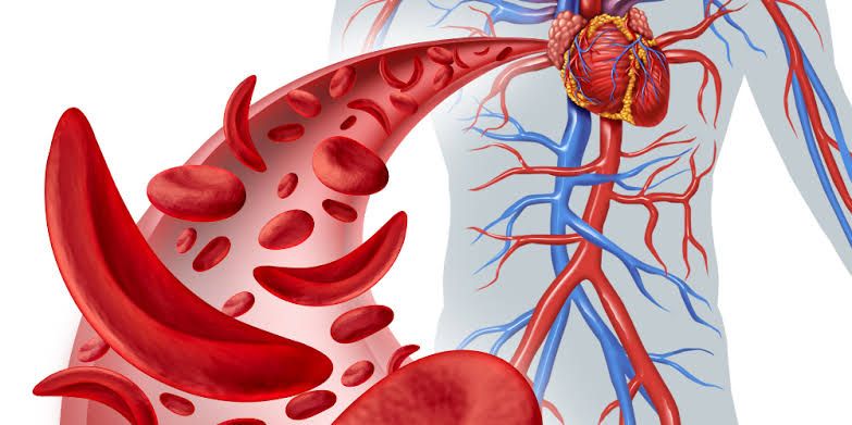
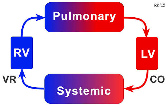
The circulatory system is divided into two main circuits: systemic circulation and pulmonary circulation. Each of these circuits has distinct characteristics, including differences in pressure that are crucial for their respective functions.
Systemic Circulation Pressures
Systemic circulation is responsible for delivering oxygenated blood from the heart to the rest of the body. The pressures involved in this circuit are significantly higher than those in pulmonary circulation due to the greater resistance encountered as blood travels through various tissues and organs.
- Left Ventricle Pressure: Blood is pumped from the left ventricle into the aorta during systole (the contraction phase). The pressure generated here can reach approximately 120 mmHg, which is necessary to overcome the high resistance of systemic arteries.
- Aortic Pressure: As blood enters the aorta, it experiences a pressure drop due to friction and resistance within the arterial walls. The normal resting pressure in the aorta is about 80 mmHg during diastole (the relaxation phase), leading to an average mean arterial pressure (MAP) of around 93-100 mmHg.
- Arterioles and Capillaries: As blood moves through smaller arteries and arterioles, pressure continues to decrease, typically dropping to about 30-40 mmHg by the time it reaches capillary beds. This lower pressure allows for efficient nutrient and gas exchange between blood and tissues.
- Venous Return: After passing through capillaries, deoxygenated blood returns through venules and veins, where pressures drop further to around 5-10 mmHg before entering the right atrium via the superior and inferior venae cavae.
Pulmonary Circulation Pressures
In contrast, pulmonary circulation carries deoxygenated blood from the right side of the heart to the lungs for gas exchange. This circuit operates under much lower pressures due to its unique anatomical structure and function.
- Right Ventricle Pressure: The right ventricle pumps deoxygenated blood into the pulmonary trunk at a peak systolic pressure of approximately 25 mmHg. This lower pressure is sufficient because pulmonary vessels have less resistance compared to systemic vessels.
- Pulmonary Arteries: Blood flows from the right ventricle into the pulmonary arteries, where pressures remain low—around 15-20 mmHg during systole and about 8-12 mmHg during diastole.
- Capillary Beds in Lungs: Within lung capillaries, pressures drop even further, typically ranging from 5-10 mmHg. This low-pressure environment facilitates gas exchange as carbon dioxide is released and oxygen is absorbed across alveolar membranes.
- Return Flow via Pulmonary Veins: Oxygenated blood returns from the lungs through pulmonary veins at similar low pressures (approximately 5 mmHg) before entering the left atrium of the heart.
Comparison of Pressures
The key difference between systemic and pulmonary circulation lies in their respective pressures:
- Systemic Circulation: High-pressure system with peak arterial pressures reaching up to 120/80 mmHg.
- Pulmonary Circulation: Low-pressure system with peak arterial pressures around 25/10 mmHg.
This disparity in pressures reflects their functional requirements; systemic circulation must deliver blood against higher resistance throughout various body tissues, while pulmonary circulation requires lower pressures suitable for gas exchange without damaging delicate lung structures.
Conclusion
Understanding these differences in pressure helps clarify how each circulatory system operates effectively within its physiological context—systemic circulation supporting metabolic demands across body tissues while pulmonary circulation facilitates efficient gas exchange within lung alveoli.
Types of Blood Flow and Significance of Reynolds Number
1. Types of Blood Flow
Blood flow in the circulatory system can be categorized into two primary types: laminar flow and turbulent flow.
- Laminar Flow: This type of blood flow occurs when blood moves in an orderly, parallel manner through the arteries. In laminar flow, the layers of blood slide smoothly past one another without disruption. This is akin to a straight river with a smooth bottom where the water flows uniformly. Laminar flow is typically observed in healthy arteries, such as the femoral artery, where there are no obstructions or irregularities that could disturb the flow pattern. The characteristics of laminar flow include:
- Smooth and constant motion.
- Minimal energy loss due to friction.
- Predominance of viscous forces over inertial forces.
- Turbulent Flow: In contrast, turbulent flow occurs when the pathway of blood becomes disorganized, leading to chaotic movement and the formation of eddy currents. This type of flow can be visualized as a winding river with varying depths and obstacles that disrupt smooth movement. Turbulent blood flow often arises in conditions where there are obstructions like atherosclerotic plaques, at points where blood vessels branch, or in areas with pronounced bends such as the ascending aorta. The characteristics of turbulent flow include:
- Disordered movement with layers breaking apart.
- Increased energy loss due to friction and chaotic eddies.
- Predominance of inertial forces over viscous forces.
2. Significance of Reynolds Number
The Reynolds number (Re) is a dimensionless quantity that plays a crucial role in predicting whether blood flow will be laminar or turbulent based on fluid dynamics principles. It is defined mathematically as:
Re = D⋅v⋅ρ ÷ μ
Where:
- D is the diameter of the vessel,
- v is the velocity of blood,
- ρ is the density of blood,
- μ is the dynamic viscosity of blood.
The significance of Reynolds number lies in its ability to indicate the nature of fluid flow:
- Laminar Flow: Occurs when Re < 2300. In this regime, viscous forces dominate, leading to smooth and orderly motion.
- Transitional Flow: Occurs when Re ranges from 2300 to 4000. This range indicates a shift between laminar and turbulent flows, where instabilities may begin to develop.
- Turbulent Flow: Occurs when Re > 4000. In this regime, inertial forces dominate, resulting in chaotic movements characterized by eddies and vortices.
Understanding these types and their relationship with Reynolds number is vital for several reasons:
- Clinical Implications: Turbulent blood flow can lead to increased vascular resistance (systemic vascular resistance), which may contribute to higher blood pressure and increased workload on the heart.
- Diagnostic Value: The presence of turbulence can produce audible sounds such as murmurs or bruits during medical examinations, aiding in diagnosing cardiovascular conditions.
- Design Considerations: Knowledge about laminar versus turbulent flows informs medical device design (e.g., stents and catheters) and surgical procedures by ensuring optimal performance under various physiological conditions.
In summary, recognizing whether blood flow is laminar or turbulent through analysis using Reynolds number has significant implications for understanding cardiovascular health and designing effective medical interventions.
Acute Local Control of Blood Flow
In physiology, acute local blood flow regulation refers to the intrinsic mechanisms that control the vascular tone of arteries at a localized level, specifically within certain tissues, organs, or organ systems. This regulation allows blood vessels to automatically adjust their diameter—either dilating (widening) or constricting (narrowing)—in response to changes in the local environment. The primary goal of this mechanism is to match the oxygen demand of tissues with the oxygen supply available in the blood.
Mechanisms of Local Blood Flow Regulation
There are two major mechanisms through which local blood flow is regulated:
- Metabolic Control: This mechanism involves metabolites and paracrine agents released from surrounding tissues that act on blood vessels. When tissue metabolism increases, it leads to a higher demand for oxygen and a decrease in available oxygen levels. This change results in a drop in pH and triggers the release of adenosine, which causes vasodilation of nearby blood vessels. For example, during physical activity, active muscles require more oxygen; thus, the blood vessels supplying these muscles will widen to increase blood flow.
- Myogenic Control: This mechanism originates from the walls of blood vessels themselves and includes muscle reflexes as well as products released from endothelial cells lining the vessels. Key substances involved include nitric oxide (NO), which promotes vasodilation, and endothelin-1, which induces vasoconstriction. These endothelial products are released in response to various stimuli such as chemical signals (e.g., histamine) or increased shear stress due to blood flow.
Examples of Local Blood Flow Regulation by Organ Type
Different organs have unique mechanisms for regulating local blood flow:
- Cerebral Circulation: The brain’s blood vessels are highly sensitive to changes in carbon dioxide levels (pCO2) and hydrogen ion concentration. An increase in pCO2 leads to vasodilation, enhancing blood flow to meet metabolic demands.
- Coronary Circulation: The heart primarily utilizes metabolic control for its local regulation through adenosine produced by neighboring cells during increased activity.
- Renal Circulation: The kidneys regulate their own blood flow through a process known as Tubuloglomerular Feedback, which adjusts renal perfusion based on filtration rates.
- Pulmonary Circulation: In contrast to other organs, pulmonary circulation exhibits hypoxic vasoconstriction; when oxygen levels are low (hypoxemia), pulmonary arteries constrict rather than dilate.
- Splanchnic Circulation: This system supplies several gastrointestinal organs and is influenced by hormones and metabolites released during digestion that promote vasodilation.
- Skeletal Muscle: Blood flow regulation here involves both metabolic control and hyperemia—an increase in blood flow due to heightened metabolic activity or after a brief interruption in circulation (reactive hyperemia).
Compensatory Mechanisms During Hypoxia
During conditions such as systemic hypoxia or local hypoperfusion (reduced blood supply), compensatory mechanisms come into play. For instance, during exercise under hypoxic conditions, there is an observed compensatory vasodilation that enhances blood flow relative to similar exercise performed under normal oxygen levels. Nitric oxide plays a significant role in this response while adenosine is particularly important during episodes of hypoperfusion.
In summary, acute local control of blood flow is essential for maintaining tissue health and function by ensuring that areas with higher metabolic needs receive adequate oxygen supply through intricate regulatory mechanisms tailored for specific organs or systems.
Acute Humoral Control of Local Blood Flow
Acute humoral control of local blood flow refers to the regulation of blood vessel diameter and, consequently, blood flow through the action of various hormones and biochemical substances present in the bloodstream. This form of control is distinct from neural regulation and intrinsic mechanisms that operate locally within tissues. The humoral response can be rapid and is crucial for adjusting blood flow to meet the metabolic demands of different organs or tissues.
Mechanisms of Acute Humoral Control
- Vasodilators and Vasoconstrictors: Various substances in the blood can cause either vasodilation (widening of blood vessels) or vasoconstriction (narrowing of blood vessels). Key vasodilators include:
- Nitric Oxide (NO): Released from endothelial cells, it diffuses into smooth muscle cells, causing relaxation and dilation.
- Adenosine: A metabolite that accumulates during increased tissue metabolism; it promotes vasodilation in response to low oxygen levels.
- Prostaglandins: These lipid compounds can also induce vasodilation in specific vascular beds.
Conversely, vasoconstrictors such as:
- Endothelin-1: A potent vasoconstrictor released by endothelial cells in response to various stimuli.
- Angiotensin II: A hormone that increases blood pressure by constricting blood vessels.
- Hormonal Influence: Several hormones play a role in regulating local blood flow through their systemic effects:
- Epinephrine/Norepinephrine: Released from the adrenal medulla, these catecholamines can cause vasodilation in skeletal muscle while inducing vasoconstriction in other vascular beds.
- Vasopressin (Antidiuretic Hormone): Increases water reabsorption in kidneys but also causes vasoconstriction, affecting overall vascular resistance.
- Local Tissue Factors: The release of certain factors from tissues can influence local blood flow:
- During periods of increased metabolic activity, tissues release metabolites like carbon dioxide (CO2), hydrogen ions (H+), and potassium ions (K+), which lead to local vasodilation.
- Inflammatory mediators such as histamine can increase local blood flow by promoting dilation and increasing permeability.
- Integration with Other Systems: Acute humoral control does not act alone; it integrates with neural mechanisms and intrinsic regulatory systems. For example, during exercise, both hormonal signals (like epinephrine) and local metabolic changes work together to enhance blood flow to active muscles while reducing it to less active areas.
- Response to Pathological Conditions: In pathological states such as shock or sepsis, acute humoral responses become critical for maintaining perfusion to vital organs. The body may release large amounts of inflammatory cytokines that alter normal vascular responses, leading to either excessive vasodilation or constriction depending on the context.
In summary, acute humoral control plays a vital role in regulating local blood flow through a complex interplay between various hormones and biochemical substances that respond dynamically to changes in tissue metabolism and systemic conditions.
Long-Term Control of Local Blood Flow
Long-term control of local blood flow is a complex physiological process that involves various mechanisms to ensure that tissues receive adequate blood supply over extended periods, particularly in response to chronic changes in metabolic demand or environmental conditions. This regulation is crucial for maintaining homeostasis and ensuring that organs function optimally.
1. Neuro-Humoral Interactions
One of the primary mechanisms involved in long-term blood flow regulation is neuro-humoral interaction. This refers to the influence of the nervous system and hormones on vascular tone and blood flow. For instance, hormones such as nitric oxide (NO) and endothelins play significant roles in modulating vascular resistance over time. Nitric oxide acts as a vasodilator, promoting increased blood flow by relaxing smooth muscle cells in the vessel walls. Conversely, endothelins are potent vasoconstrictors that can reduce blood flow by causing the smooth muscles to contract.
2. Paracrine Signaling
Paracrine signaling involves the release of signaling molecules from nearby cells that affect local blood vessels. These substances can include metabolites produced during cellular metabolism, which signal for increased blood flow when tissue demands rise. For example, during prolonged exercise or sustained activity, tissues may release adenosine and other vasodilators that promote increased perfusion to meet elevated oxygen requirements.
3. Autoregulation Mechanisms
Autoregulation refers to the ability of an organ to maintain a relatively constant blood flow despite changes in perfusion pressure. This mechanism is particularly important for organs like the kidneys and brain, where consistent blood supply is critical for function. In autoregulation, myogenic responses (where smooth muscle cells react directly to changes in stretch due to pressure) help adjust vessel diameter accordingly.
4. Long-Term Adaptations
Over longer periods, chronic changes in local blood flow can lead to structural adaptations within the vascular system itself. For example, if a tissue consistently experiences higher metabolic demands (such as skeletal muscle during regular exercise), it may undergo angiogenesis—the formation of new blood vessels—to enhance its capacity for oxygen delivery and nutrient exchange.
5. Renal Regulation
The kidneys play a pivotal role in long-term regulation of systemic and local blood flow through mechanisms like tubuloglomerular feedback, which adjusts renal blood flow based on sodium chloride concentration detected by specialized cells within the nephron. This feedback loop helps maintain stable glomerular filtration rates and overall fluid balance.
In summary, long-term control of local blood flow involves a combination of neuro-humoral interactions, paracrine signaling from metabolically active tissues, autoregulatory mechanisms inherent to specific organs, structural adaptations such as angiogenesis, and renal regulatory functions that collectively ensure that each organ receives adequate perfusion according to its needs over time.
Auto Regulation of Blood Flow in Organs
Auto regulation of blood flow refers to the ability of certain organs to maintain a relatively constant blood flow despite changes in arterial pressure. This is primarily achieved through two mechanisms: metabolic and myogenic mechanisms. The following organs are known for their auto regulatory capabilities:
1. Kidneys
The kidneys exhibit strong auto regulation, primarily through the myogenic mechanism. When there is an increase in arterial pressure, the smooth muscle cells in the afferent arterioles respond by constricting, which helps prevent excessive blood flow into the glomeruli and maintains glomerular filtration rate (GFR). Conversely, during low arterial pressure, these arterioles dilate to allow more blood flow.
2. Brain
The brain also has a robust auto regulatory system that ensures adequate cerebral perfusion. Metabolic mechanisms play a significant role here; for instance, when carbon dioxide levels rise or pH decreases (indicating increased metabolic activity), cerebral vessels dilate to enhance blood flow. Myogenic responses are also present; if arterial pressure increases, cerebral arteries constrict to protect against potential damage from high pressures.
3. Heart
The coronary circulation demonstrates auto regulation through both metabolic and myogenic mechanisms. During periods of increased myocardial oxygen demand (such as during exercise), metabolites like adenosine accumulate, leading to vasodilation of coronary arteries. Additionally, if there is an increase in perfusion pressure, coronary vessels can constrict to prevent excessive blood flow.
4. Skeletal Muscle
Skeletal muscles utilize metabolic auto regulation during physical activity. When muscles contract and metabolize glucose for energy, they produce metabolites such as lactic acid and adenosine that cause vasodilation of local arterioles, increasing blood flow to meet the heightened demand for oxygen and nutrients.
5. Lungs
In pulmonary circulation, auto regulation occurs differently compared to systemic circulation due to the unique function of the lungs in gas exchange. The myogenic response is evident; when pulmonary arterial pressure rises, pulmonary arterioles constrict (a response known as hypoxic pulmonary vasoconstriction) to redirect blood flow away from poorly ventilated areas of the lung.
6. Intestines
The intestines also exhibit auto regulation primarily through metabolic mechanisms. After a meal, increased nutrient absorption leads to elevated levels of metabolites that cause vasodilation in mesenteric vessels, enhancing blood supply for digestion and absorption.
In summary, the primary organs where auto regulation of blood flow occurs include the kidneys, brain, heart, skeletal muscle, lungs, and intestines. Each organ employs a combination of metabolic and myogenic mechanisms tailored to its specific physiological needs.


